Loading the model
This device cannot show the 3D model. You can read the guide steps below as text.
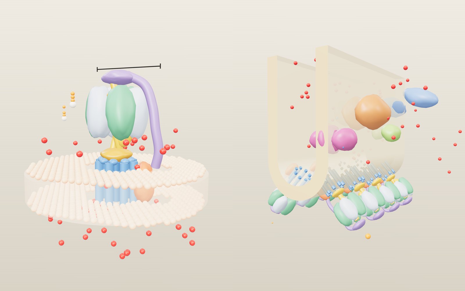
1 / 17 A machine in the membrane This is a simplified ATP synthase in the inner membrane of a mitochondrion. The head on top is 10 nm across; the bar above it gives the scale. The shapes are smooth blobs, not measured surfaces. The bar at the bottom of the stage opens five more scenes.
Drag to turn. Scroll or pinch to zoom.
Guide Parts Controls
01 A machine in the membrane This is a simplified ATP synthase in the inner membrane of a mitochondrion. The head on top is 10 nm across; the bar above it gives the scale. The shapes are smooth blobs, not measured surfaces. The bar at the bottom of the stage opens five more scenes.
02 Protons pile up on one side The respiratory chain has pumped protons below the membrane. Two things push them back: the charge difference Δψ and the pH difference ΔpH. Together they make the proton-motive force, Δp = Δψ − 59·ΔpH, shown at the top of the readout.
03 Each proton turns the ring a little A proton enters the a subunit, lands on the acidic side chain of a c subunit and rides once round before leaving on the matrix side. With 8 c subunits, 8 protons turn the ring once.
04 An arm holds the head still If the head turned with the stalk, nothing would change inside it. The peripheral stalk runs from the a subunit to the top of the head and keeps the α and β subunits in place.
05 The axle turns inside the head The central stalk is fixed to the c ring, so it turns with it. The head is cut open here: watch the bulge on the stalk sweep past the β subunits one after another.
06 Three sites, three states Seen from the matrix side, each β is open, loose or tight; the colours show which. ADP and phosphate bind in the loose site, become ATP in the tight site and leave when it opens. One turn makes three ATP.
07 Where the rotor stalls The rotor only turns forward if the protons in one turn bring more energy than three ATP cost. With c8 and 50 kJ/mol that balance falls at about 194 mV, and here the rotor stands still. The dashed line in the chart marks it.
08 When ATP drives it the other way Lower the gradient below the stall point and ATP wins: the stalk turns backwards in 120° steps and the ring pumps protons out. The counter for ATP used goes up.
09 Drugs that stop it Oligomycin wedges into the c ring next to the a subunit, so no proton can get on and the rotor stops. Try the other inhibitors in Controls: DCCD, bedaquiline, the uncoupler DNP and the brake IF1.
10 Where the protons come from Inside a mitochondrion the inner membrane folds into cristae. Complexes I, III and IV take electrons from NADH down to oxygen and pump ten protons for every two electrons into the fold.
11 Pairs on the ridge ATP synthase sits in V-shaped pairs along the edge of each fold. The protons come back through them; switch Fuel to succinate and see the P/O ratio fall from 2.7 to 1.6.
12 Light instead of food In a chloroplast, light splits water at photosystem II and the chain fills the thylakoid lumen with protons. The chloroplast enzyme has a c14 ring, so it needs 4.67 protons per ATP.
13 A bacterium uses the same gradient E. coli pumps protons out across its inner membrane. The same gradient drives ATP synthase and the flagellar motor, shown side by side at the same scale. Switch Flagella clockwise to make the cell tumble.
14 Built to run backwards A V-type ATPase uses ATP to pump protons into a lysosome until the inside reaches pH 4.5 to 5. It has the same kind of rotor and head, with three peripheral stalks.
15 Seeing it turn In 1997 Noji and colleagues stuck F1 upside down on glass and tied a fluorescent actin filament to γ. With ATP the filament turned anticlockwise, seen from the membrane side. Longer filaments turn more slowly.
16 One ATP, one step With very little ATP the filament waits for each molecule, then jumps 120°. Torque 40 pN·nm times 120° is about 84 pN·nm of work, close to the energy in one ATP.
17 Turning it by hand With a magnetic bead on γ, electromagnets can turn F1 backwards. Itoh and colleagues showed in 2004 that this makes ATP: the enzyme runs as a synthase when it is turned the right way.
Inner membrane The inner membrane of a mitochondrion. The respiratory chain pumps protons from the matrix (above) into the intermembrane space (below), and ATP synthase lets them back. Protons (H⁺) Red beads. The more crowd below the membrane, the larger the proton-motive force. A proton that crosses rides once round the c ring on one c subunit. c ring A ring of identical c subunits. Each is a hairpin of two helices with an acidic side chain halfway up (dark bead). Eight in cow and human mitochondria, 9 to 15 in bacteria and chloroplasts. a subunit The fixed part of Fo beside the ring, made from the mitochondrial gene MT-ATP6. Protons enter through one half-channel, ride the ring and leave through the other. Peripheral stalk The long arm from the a subunit to the cap on top of the head. It stops the α and β subunits turning with the central stalk. Central stalk (γ) The axle. Its foot is fixed to the c ring and its coiled part reaches up into the head; a bulge on one side presses on the β subunits in turn. α subunits Three subunits between the β subunits. They bind nucleotides but do not make ATP. β subunits The three subunits that make ATP. At any moment one is open (O), one loose (L) and one tight (T); each 120° turn moves all three on by one state. ADP, Pi and ATP White bead for adenosine, orange beads for phosphates. ADP and phosphate bind in a loose site, become ATP in the tight site and leave when it opens. Inhibitor The chosen inhibitor: oligomycin or bedaquiline on the c ring, DCCD on the proton site, DNP leaking protons through the lipid, or IF1 wedged into the head. Crista A fold of inner membrane reaching into the matrix. The space inside the fold is joined to the intermembrane space, so pumped protons collect there. Complex I NADH dehydrogenase, an L-shaped enzyme of 45 subunits. It takes two electrons from NADH, passes them to ubiquinone and pumps four protons. Complex II Succinate dehydrogenase, also part of the citric acid cycle. It feeds electrons from succinate to ubiquinone without pumping protons. Ubiquinone (Q) A small fat-soluble carrier that moves inside the membrane, taking electrons from complex I or II to complex III. Complex III Cytochrome bc1, a dimer. It passes electrons from ubiquinol to cytochrome c and releases four protons on the outer side per two electrons. Cytochrome c A small protein on the outer face of the membrane that carries one electron at a time from complex III to complex IV. Complex IV Cytochrome c oxidase. It hands electrons to oxygen, making water, and pumps two protons per two electrons. ATP synthase dimers Pairs of ATP synthase, 86° apart, in rows along the ridge of the crista. The pairs help bend the membrane. Thylakoid membrane The membrane inside a chloroplast. Light reactions fill its lumen with protons; the stroma outside is where ATP and NADPH are used. Photosystem II A dimer with its light-harvesting antenna. Light drives electrons out of water: 2 H₂O gives O₂ and four protons in the lumen. Cytochrome b6f Passes electrons from plastoquinol to plastocyanin and moves extra protons into the lumen. Plastocyanin A small copper protein in the lumen that carries electrons from cytochrome b6f to photosystem I. Photosystem I A second light-driven step that lifts electrons to ferredoxin and on to NADP⁺, making NADPH in the stroma. Chloroplast ATP synthase CF1Fo with a c14 ring, head in the stroma. A redox switch in its γ subunit locks it in the dark. E. coli cell A rod-shaped bacterium about 2 µm long. Its inner membrane does the job of the mitochondrial inner membrane. Flagella Helical filaments turned by motors at their base. Anticlockwise they bundle and the cell swims; clockwise they fly apart and the cell tumbles. Flagellar motor About 45 nm across. Protons flowing through the stator units around it turn the rotor; CheY-P binding to the C ring switches the direction. E. coli ATP synthase The bacterial enzyme with a c10 ring. Without oxygen it can run backwards on ATP to keep the gradient up. Respiratory enzymes NADH dehydrogenase and the bo3 oxidase in the inner membrane of E. coli. They pump protons out into the periplasm. Lysosome A small digestive compartment. Its inside is kept at pH 4.5 to 5; the colour of the small sphere follows the lumen pH. V-ATPase A rotary proton pump driven by ATP: V1 in the cytosol, Vo with its proteolipid ring in the membrane, three peripheral stalks. V1 head The ATP-splitting head. When yeast is starved of glucose, V1 lets go of Vo and pumping stops. Chloride exchanger ClC-7 lets chloride in while protons go out, so charge does not build up and the pump can keep acidifying. Coverslip The glass the F1 molecules are stuck to by histidine tags on their β subunits. The microscope looks at it from above. F1 on glass α3β3γ upside down: head on the glass, γ sticking up. Enlarged here; in the main view it is too small to see. Probe What shows the turning: a fluorescent actin filament (1997), a 40 nm gold bead (2001) or a magnetic bead (2004). Electromagnets Four coils that make a rotating field. They turn the magnetic bead, and with it γ, backwards so that F1 makes ATP.