FIG. 43How It Works
How ATP synthase works
In mitochondria the rotor of ATP synthase turns about 100 times a second, and every full turn releases three molecules of ATP.
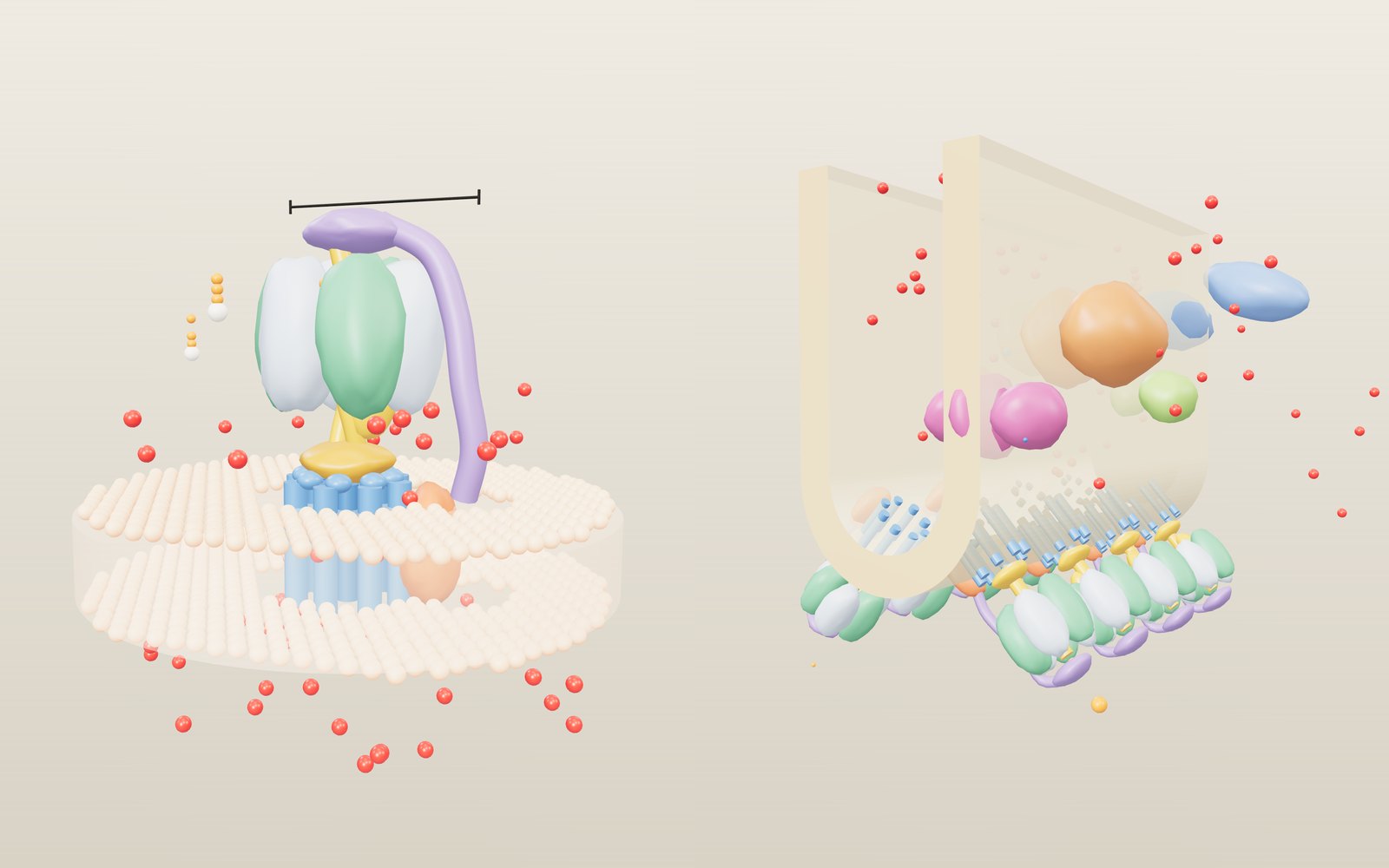
This device cannot show the 3D model. You can read the guide steps below as text.
1 / 17 A machine in the membraneThis is a simplified ATP synthase in the inner membrane of a mitochondrion. The head on top is 10 nm across; the bar above it gives the scale. The shapes are smooth blobs, not measured surfaces. The bar at the bottom of the stage opens five more scenes.
Drag to turn. Scroll or pinch to zoom.
Where does the ATP in a cell come from?
How does ATP synthase work? Protons flowing back across a membrane turn a ring in the membrane like a water wheel, and the turning stalk squeezes the three catalytic sites in the head one after another, joining ADP and phosphate into ATP. Run backwards, the same enzyme splits ATP and pumps protons, which is why it is also called an ATPase.
Muscles, nerves and the enzymes that copy DNA all run on the same fuel, adenosine triphosphate. Cut off its third phosphate and the energy released drives the work; join it back on and the molecule is ready again. The amounts are large. According to the Nobel Committee, an adult at rest turns over roughly half of their body weight in ATP each day, and during hard work the figure can approach a tonne. A cell keeps only a small stock, so the ATP has to be rebuilt as fast as it is spent.
Most of that rebuilding is done by one enzyme, ATP synthase. It sits in the inner membrane of mitochondria, in the membranes of bacteria and in the thylakoids of chloroplasts. It is also one of the few proteins that work by turning. This exhibit builds a simplified version of it at molecular scale and then follows it into five other places: a fold of mitochondrial membrane with the respiratory chain, a chloroplast membrane in the light, a swimming bacterium, a lysosome where a related pump runs on ATP, and the glass slide of the 1997 experiment that first showed the rotor turning. The bar at the bottom of the stage switches between the six scenes.
A dam made of membrane
The energy does not come from food directly. Respiration passes electrons from food down a chain of membrane proteins, and that chain uses their energy to pump protons out of the matrix (the inside of the mitochondrion) into the narrow space between its two membranes. Protons build up on one side. The result is a difference in concentration and in electric charge across the membrane, which together are called the proton-motive force.
Peter Mitchell proposed this chemiosmotic idea in 1961 and received the Nobel Prize in Chemistry for it in 1978. It means a cell stores energy the way a dam does: as a difference in level that wants to even out. ATP synthase is the turbine in the dam. Protons can flow back into the matrix only through it, and as they pass they turn its rotor.
In the model, protons are the red beads. The two sliders Membrane potential and pH difference set how hard they push. Move either one and the crowd below the membrane grows or thins, and the rotor speeds up, slows down, stops or turns the other way.
Adding up the proton-motive force
A proton outside the matrix is pushed inward for two reasons. The matrix is negatively charged compared with the outside, and it has fewer protons, so its pH is higher. Mitchell wrote the total push as one number, the proton-motive force Δp, in millivolts:
Δp = Δψ − 59 × ΔpH
Here Δψ is the electrical potential of the matrix minus that of the outside, and ΔpH is the pH of the matrix minus that of the outside. In a working mitochondrion Δψ is negative and ΔpH positive, so both terms make Δp more negative, and the more negative it is, the harder protons push in. The factor 59 converts one pH unit into millivolts: it is ln 10 × RT/F, which comes to 59.16 mV at 25 °C and 61.5 mV at body temperature, 37 °C. The model uses 59.
With the sliders at their starting values, Δψ is −150 mV and ΔpH is 0.80, so Δp = −150 − 59 × 0.80 = −197 mV. The readout writes out this sum each time you move a slider. Brand and Nicholls give 150 to 180 mV as typical sizes for the membrane potential of mitochondria in cells; in mitochondria most of the push is electrical. In chloroplasts it is the other way round, and in strong light most of it is stored as a pH difference across the thylakoid membrane.
What matters for the enzyme is the energy each proton brings. One mole of protons crossing 197 mV releases F × Δp = 96,485 × 0.197 ≈ 19 kJ. The rest of the exhibit comes back to this number again and again.
Fo, the part in the membrane
The enzyme has two halves with historical names. Fo, from oligomycin-sensitive factor, is the part in the membrane. F1, factor 1, is the knob that sticks out into the matrix. Efraim Racker's group isolated the complete enzyme from mitochondria in 1960.
The moving part of Fo is the c ring, a ring of identical small proteins. Each c subunit is a hairpin of two helices, and halfway up the outer helix sits an acidic side chain (a glutamate in mitochondria, shown as a dark bead). Next to the ring is the a subunit, which does not turn. It offers a proton two half-channels: one opens to the space outside, the other to the matrix.
The trick is that a charged side chain cannot face the oily inside of the membrane. A c subunit can only turn away from the a subunit after a proton has landed on its glutamate and neutralised it. So a proton comes in through the first half-channel, rides once round the ring on its c subunit, and leaves through the second half-channel into the matrix. Because there are more protons outside than inside, they arrive more often on one side than the other, and the ring turns in one direction. Pogoryelov and colleagues showed in crystal structures of a c15 ring how the site stays locked shut while it faces the lipid and opens only at the a subunit. In the model, the beads on the ring are protons on their way round.
F1, the part that makes ATP
F1 is a ring of six subunits, three α and three β, alternating like segments of an orange. Noji and colleagues described it as a barrel about 5 nm in radius, which is why the head in the model is drawn 10 nm across. The scale bar above the head marks those 10 nm.
Only the β subunits make ATP. Up the middle runs the central stalk, built mainly from the γ subunit, whose rotor part is only about 1 nm in radius. Its foot is fixed to the c ring, so the stalk turns with the ring. On the other side, a long peripheral stalk runs from the a subunit up to a cap on top of F1 and holds the α3β3 head still. Without that arm the head would simply turn with the axle and nothing inside would change.
The γ subunit is not symmetrical. One side bulges, and as it turns it presses on each β subunit in turn and changes its shape. Switch on Cut away the head and the half of the head facing you disappears, so you can watch the bulge sweep past.
Three sites take turns
Over several decades of isotope experiments, Paul Boyer found something unusual about this enzyme. Joining ADP and phosphate inside the enzyme takes little energy. The energy is spent on binding ADP and phosphate tightly enough and, above all, on letting go of the finished ATP. He proposed that the three β sites take turns and pass through three states:
- Open (O): the site is empty or just releasing an ATP.
- Loose (L): ADP and phosphate bind loosely.
- Tight (T): the site closes, and ADP and phosphate become ATP.
Each 120° turn of the stalk moves every site one state on. The tight site opens and releases its ATP, the loose one closes, and the open one picks up new ADP and phosphate. Boyer called this the binding change mechanism, and he proposed that the stalk turns. Switch on Colour β by state and the three β subunits turn pale for open, yellow for loose and red for tight; the readout writes the same states as letters. In the Guide step seen from above you can follow ADP (white and orange beads) and a separate phosphate becoming one ATP before it flies off.
John Walker approached the same enzyme from the structure. With Jan Pieter Abrahams and Andrew Leslie he solved the crystal structure of F1 from cow heart at 2.8 Å in 1994. The three β subunits really were in three different shapes with different nucleotides bound, and the asymmetric γ sat in the middle. The structure fitted Boyer's mechanism. Boyer and Walker shared half of the 1997 Nobel Prize in Chemistry; the other half went to Jens Skou for the sodium-potassium pump, another membrane enzyme that runs on ATP. The Nobel Committee notes that at rest that pump alone uses about a third of all the ATP made.
Where the rotor stalls
Why does the rotor turn forward at all? Count the energy. One full turn moves n protons across the membrane, where n is the number of c subunits, and makes three ATP. So each ATP gets n/3 protons' worth of energy:
energy in per ATP = (n/3) × F × Δp
Making an ATP inside a cell costs a definite amount too, the phosphorylation potential, written ΔGp. It depends on how much ATP, ADP and phosphate are around; the model starts at 45 kJ/mol, and the Cost of one ATP slider changes it. If the energy in is larger than the cost, the rotor turns forward and makes ATP. If it is smaller, ATP wins and pushes the rotor backwards. Where the two are equal, the rotor stands still:
Δp at stall = 3 × ΔGp / (n × F)
For the cow ring, n = 8, at 50 kJ/mol that is 3 × 50,000 / (8 × 96,485) = 0.194 V, or 194 mV. At 45 kJ/mol it is 175 mV. The chart in the readout plots the model's rotor speed against Δp for the ring and cost you have chosen, with a dashed line at the stall point and a dot where your sliders are. The table under the controls does the same sum for every ring size from c8 to c15.
Two things follow. A bigger ring stalls at a lower gradient: c15 needs only 93 mV to balance the same 45 kJ/mol, because it brings 15 protons per turn instead of 8. And the enzyme is reversible. Nothing in it decides which way it turns; the balance of the two energies does. The speed rule in the model (a smooth curve through the stall point) is made up for the picture, but the position of the stall point is calculated.
Running backwards
Lower the gradient below the stall point, or raise the cost of an ATP, and the readout switches to Using ATP, pumping protons. Now ATP binds in the head, is split, and the stalk turns the other way in 120° steps. The c ring carries protons from the matrix out across the membrane. The counter for ATP used goes up.
In a healthy mitochondrion this rarely happens, because the respiratory chain keeps the gradient high. When it collapses, for example when oxygen runs out, ATP synthase could drain the cell's ATP in a few minutes by running backwards. Mitochondria carry a brake for this: a small protein called IF1. Cabezón and colleagues showed in 2003 that it wedges into one α-β interface of F1 and touches γ. In the model, choose IF1 as the Inhibitor and push the gradient down: the rotor stops instead of turning backwards, and the helix glows while it holds.
Bacteria put the backwards mode to use. Without oxygen, E. coli makes ATP by fermentation and then lets ATP synthase run in reverse to keep a proton gradient, which it needs for its flagella and for moving nutrients across the membrane. The Bacterium scene shows this when you switch Oxygen off. Some relatives were built for the backwards job from the start; they appear in the V-ATPase scene.
Drugs and poisons that stop it
Because so much depends on one enzyme, several natural and man-made molecules target it. The Inhibitor control shows five of them.
- Oligomycin gave Fo its name. Symersky and colleagues solved it bound to the yeast c10 ring in 2012: it sits on the outside of the ring between two c subunits and covers the glutamate that carries the proton, so no proton can get on and the rotor stops. The residues that form its pocket are the same in yeast and humans. Laboratories use it to measure how much respiration goes into ATP and how much leaks away.
- DCCD (dicyclohexylcarbodiimide) reacts with that same carboxylate and blocks it permanently. Pogoryelov and colleagues used DCCD-modified c15 rings to show how the proton site opens and closes. In the model the glutamate beads turn green.
- Bedaquiline is a tuberculosis drug. Andries and colleagues found it in 2005 as a compound that kills Mycobacterium tuberculosis; resistant mutants changed the proton-carrying part of ATP synthase. In 2015 Preiss and colleagues solved it on a mycobacterial c9 ring: it covers the ion sites so the ring cannot carry protons. They describe it as the first drug in 40 years approved for multidrug-resistant tuberculosis. A 2024 study showed it can bind the human enzyme as well, at a similar place on the c ring.
- DNP (2,4-dinitrophenol) does not touch the enzyme. It carries protons through the lipid itself, so the gradient collapses and food is burned as heat without making ATP. In 1933 it was sold as a slimming drug; it causes dangerous overheating, and a 2011 review counted 62 published deaths. In the model the protons cross straight through the membrane and the rotor stalls. Brown fat does a controlled version of the same thing with its own protein, UCP1, to keep animals warm.
- IF1, the cell's own brake, stops only the backwards direction, as described above.
Rings of different sizes
The size of the c ring sets the price of an ATP. With n subunits it takes n/3 protons per ATP. Watt and colleagues found 8 c subunits in cow heart mitochondria in 2010, and pointed out that the c subunit sequence is identical in almost all vertebrates, so very probably every vertebrate pays 2.7 protons per ATP. Fungi, bacteria and chloroplasts use rings of 9 to 15. The table under the controls lists 13 rings and the structures that show them:
| Organism | Ring | H⁺ per ATP | Structure |
|---|---|---|---|
| Cow heart mitochondria | c8 | 2.67 | PDB 2XND |
| Mycobacterium phlei | c9 | 3.00 | PDB 4V1F |
| Yeast | c10 | 3.33 | PDB 1QO1 |
| Escherichia coli | c10 | 3.33 | PDB 5T4O |
| Ilyobacter tartaricus (Na⁺) | c11 | 3.67 | PDB 1YCE |
| Bacillus pseudofirmus OF4 | c13 | 4.33 | PDB 2X2V |
| Spinach chloroplast | c14 | 4.67 | PDB 2W5J |
| Spirulina platensis | c15 | 5.00 | PDB 2WIE |
Why not use the smallest ring everywhere? A small ring makes ATP cheaply but needs a high gradient to reach its stall point; a large ring works with a weaker push but spends more protons. Bacillus pseudofirmus OF4 lives in alkaline water, where protons outside the cell are scarce, and its c13 ring shares each proton with a bound water molecule. Ilyobacter tartaricus goes further and carries sodium ions instead of protons. Vollmar and colleagues found that the ion sites on the spinach c14 ring are 10.6 to 10.8 Å apart, the same spacing as on the Ilyobacter c11 ring, so a ring with more subunits is simply wider.
Set c ring size on the Enzyme scene and watch the stall point and the cost per ATP move together. The ring slider names the measured organism for the sizes where one exists; c12 has no entry in the table.
Inside a mitochondrion
Open the Mitochondrion scene. The inner membrane of a mitochondrion is folded into ridges called cristae; the scene shows one fold in cross-section, cut open at the front. Outside the fold is the matrix. Inside the fold is a narrow space that joins the space between the two membranes, and that is where the pumped protons collect.
Four complexes on the side of the fold pass electrons from food to oxygen:
- Complex I takes two electrons from NADH and passes them to ubiquinone, a small carrier that moves inside the membrane. It is one of the largest enzymes in the cell: Zhu, Vinothkumar and Hirst placed all 45 of its subunits in the cow enzyme in 2016. It pumps 4 protons for every two electrons.
- Complex II feeds electrons from succinate into the same ubiquinone pool without pumping any protons. It is also part of the citric acid cycle.
- Complex III, cytochrome bc1, passes the electrons from ubiquinol to cytochrome c and releases 4 protons on the outer side for every two electrons.
- Complex IV, cytochrome c oxidase, hands them to oxygen, which becomes water, and pumps 2 more protons.
So two electrons from NADH move 10 protons, while two from succinate move only 6. Watt and colleagues used these numbers to work out the yield. Making one ATP with a c8 ring takes 8/3 = 2.67 protons, and getting it out of the mitochondrion costs one more: the carrier that swaps ATP for ADP and the one that brings in phosphate together use the equivalent of one proton. That makes 3.67 protons per ATP delivered. Then:
- NADH: 10 / 3.67 = 2.7 ATP per oxygen atom
- succinate: 6 / 3.67 = 1.6 ATP per oxygen atom
These are the P/O ratios. Hinkle's review of measurements gives about 2.5 and 1.5, a little below because some protons leak back through the membrane without passing the enzyme. The readout shows the calculated ratio and the one counted in the model as it runs. Switch Fuel to succinate and both drop.
The ATP synthases in this scene sit in pairs on the ridge of the fold. Davies and colleagues found with electron cryotomography of yeast mitochondria that the pairs form V shapes with an angle of 86° between them and line up in rows along the ridges. Yeast strains that cannot make the pairs have no proper cristae, only balloon-shaped ones or none. Guo, Bueler and Rubinstein's atomic model of the paired Fo region shows how the pair bends the membrane. In the model the pairs are four V shapes along the ridge, each copy turning its own rotor.
The scene runs a small proton circuit. The chain pumps faster when the gradient is low and slows as it rises (respiratory control); ATP synthase and a small leak let protons back. Try the inhibitors here too. Oligomycin stops ATP synthase, the gradient rises and respiration almost stops. DNP opens a leak, the gradient falls, respiration races, and the fuel turns into heat; the readout then shows the enzyme turning backwards on ATP, which is what uncoupled mitochondria do unless IF1 stops them. Switch Oxygen off and the whole chain stops.
Inside a chloroplast
The Chloroplast scene shows a patch of thylakoid membrane. The stroma is above, the lumen (the inside of the thylakoid) below. Here the energy comes from light, not food, but the enzyme is the same kind, and its head faces the stroma.
- Photosystem II absorbs light and pulls electrons out of water. Umena and colleagues' 1.9 Å structure shows the Mn₄CaO₅ cluster where this happens: two water molecules give one O₂, four electrons and four protons, released into the lumen.
- Plastoquinone carries the electrons through the membrane to cytochrome b6f, which moves more protons into the lumen.
- Plastocyanin, a small copper protein in the lumen, carries them to photosystem I, which uses a second photon to lift them to ferredoxin and NADP⁺. NADPH forms in the stroma.
Kramer and Evans count 12 protons into the lumen and 2 NADPH for every O₂ in this linear flow. The chloroplast enzyme has a c14 ring, so it needs 14/3 = 4.67 protons per ATP, and 12 protons make 12 × 3 / 14 = 2.57 ATP. That is 1.29 ATP per NADPH, but the Calvin cycle uses 3 ATP for every 2 NADPH, 1.5 per NADPH. The shortfall is made up by other routes, such as cyclic electron flow around photosystem I, which pump protons without making NADPH. The readout shows both ratios and recalculates them if you change the ring.
The lumen becomes acidic. In living Arabidopsis leaves Takizawa and colleagues estimated it at about pH 7.5 in weak light and about pH 5.7 in strong light with little CO₂. The chart in the readout follows the lumen pH as the light changes. In the model the stroma is fixed at pH 8 and 70 % of the gradient is stored as ΔpH; both are model choices.
Turn the light down to zero. After a short time the readout says the γ switch has locked the enzyme. Hahn and colleagues' 2018 structure of the complete chloroplast enzyme, 26 subunits, shows a β-hairpin in γ that acts as a redox switch and blocks rotation in the dark.
The chloroplast enzyme also starred in a classic experiment. In 1966 André Jagendorf and Ernest Uribe soaked spinach thylakoids in acid, moved them into an alkaline buffer with ADP and phosphate, and got ATP in the dark. A pH difference alone was enough, as Mitchell's idea required.
A bacterium and its flagella
Bacteria have no mitochondria. Their respiratory chain sits in the cell membrane itself and pumps protons out into the periplasm, the thin space between the inner and outer membranes. The Bacterium scene shows an E. coli cell about 2 µm long at the back right and, in front, a patch of its inner membrane drawn about 50 times larger than the cell.
Two machines in that patch use the same gradient. One is ATP synthase. Fillingame's group showed in 2001 that E. coli prefers a c10 ring, and Sobti and colleagues' cryo-EM structure shows the ten subunits and an ε subunit that can stretch out to block the enzyme. The other is the flagellar motor. Howard Berg describes it as about 45 nm across and built from about 20 kinds of parts; here it is drawn at the same scale as ATP synthase, more than four times wider. Protons flowing through a ring of stator units around its edge turn the rotor, and Meister, Lowe and Berg showed that the proton flux is proportional to the motor's speed.
Unlike ATP synthase, this motor reverses on command. When a protein called CheY carries a phosphate, it binds to the C ring at the bottom of the motor and switches it from counterclockwise to clockwise. In E. coli, counterclockwise rotation lets the filaments wind into a bundle and the cell swims; clockwise rotation breaks the bundle up and the cell tumbles and sets off in a new direction. Switch on Flagella clockwise: the pink CheY beads bind, the filaments fly apart and the cell tumbles.
Switch Oxygen off. Respiration stops, the gradient falls below the stall point of the c10 ring, and ATP synthase starts to run backwards on ATP from fermentation, pumping protons out to keep the motor turning. The gradient settles a little lower and the cell keeps swimming, more slowly.
Pumps built to run backwards
Some relatives of ATP synthase spend ATP as their normal job. The vacuolar or V-type ATPases pump protons into compartments of the cell to make them acidic. Lysosomes, the cell's digestive bags, are kept between pH 4.5 and 5, where their enzymes work. Mindell's review explains that the pump cannot do it alone: as protons go in, charge builds up, so another ion has to move too. A chloride/proton exchanger, ClC-7, is part of the answer, and the scene shows it beside the pumps.
The V-ATPase scene shows two pumps on a patch of lysosome membrane, with the whole lysosome small at the back; its colour follows the pH inside. The parts look familiar: a head of three A and three B subunits (the counterparts of β and α), a central rotor and a ring in the membrane. The differences are visible too. Zhao, Benlekbir and Rubinstein's cryo-EM of the yeast enzyme shows three peripheral stalks instead of one and a ring built from more than one kind of proteolipid; Mazhab-Jafari and colleagues later identified them as c8, c′ and c″. Murata and colleagues solved the ten-subunit rotor of a related sodium pump from Enterococcus hirae.
The V-ATPase also has a switch the F-type enzyme lacks: its two halves can come apart. When yeast is starved of glucose, the V1 head lets go of the Vo ring and pumping stops. Mazhab-Jafari and colleagues used exactly this to get the free Vo ring and build its atomic model. Switch on Starve: V1 lets go, the heads lift off and the lumen slowly drifts back towards neutral.
Seeing it turn
Rotation was still an inference until 1997. Hiroyuki Noji, Ryohei Yasuda, Masasuke Yoshida and Kazuhiko Kinosita fixed F1 to a glass surface by its β subunits, upside down, attached a fluorescent actin filament to the γ subunit and added ATP. Under the microscope the filament turned for more than 100 revolutions, always anticlockwise when viewed from the membrane side. F1 alone does not make ATP; given ATP, it breaks it down and turns the stalk the opposite way from synthesis.
The Rotation test scene rebuilds this. The main view is what the microscope saw: the glass, and a green filament spinning about one end. The small picture beside it shows F1 itself, drawn about 50 times larger, with γ turning in the fixed head.
The filament is not driven at a set speed. The motor pushes with a torque of about 40 pN·nm, and the water drags on the filament. For a rod turning about one end, the drag grows with the cube of its length:
drag = (4π/3) η L³ / (ln(L / 2r) − 0.447)
where η is the viscosity of water and r the radius of the filament. A 1 µm filament comes out at about 6.3 turns a second, a 2 µm one at under one turn a second. Change Actin length and the readout recalculates it; the label "drag of the probe" means the filament, not the enzyme, sets the speed.
Later work used smaller markers. Yasuda and colleagues found in 1998 that at low ATP the filament moves in discrete 120° steps, one per ATP, and that the work done in each step stays about the same across a broad range of loads, close to the free energy of hydrolysing one ATP. With a 40 nm gold bead, light enough not to slow the motor, they split each step in 2001 into substeps of roughly 90° and 30°, and measured about 130 revolutions per second when ATP was plentiful. Sakaki and colleagues found the same torque of about 40 pN·nm at 2 mM, 60 nM and 2 nM ATP, and a binding rate of 2 × 10⁷ per molar per second.
The model uses those numbers. Each ATP binds after a random wait whose average is 1 / (kon × [ATP]); then the probe swings through 120° as fast as the torque can drag it. Lower ATP concentration to around 0.2 µM and the waits become longer than the swings: the filament now jumps, and the chart draws a staircase. Choose the gold bead and the steps become sharp, because the bead hardly slows the motor; the readout then says ATP binding sets the speed, and the playback is slowed so you can follow it.
The last step reversed the experiment. Itoh and colleagues attached a magnetic bead to γ and turned it backwards with electromagnets in 2004. ATP appeared in the solution. Rondelez and colleagues shut single F1 molecules in chambers a few femtolitres in size and found that up to 77 % of the forced turns produced ATP once the small ε subunit was present. Choose Magnetic bead and switch on Magnets turn it back: the coils light up in turn, the bead goes clockwise and every 120° adds one ATP to the counter.
Checking the numbers
Each c subunit carries one proton per turn, and each turn makes three ATP. So the number of protons per ATP is the number of c subunits divided by 3: 8 ÷ 3 = 2.67 for the cow ring, 10 ÷ 3 = 3.33 for yeast and E. coli, 14 ÷ 3 = 4.67 for spinach and 15 ÷ 3 = 5.00 for Spirulina platensis.
Watt and colleagues give the rotor speed in mitochondria as about 100 turns per second. At 3 ATP per turn that is about 300 ATP per second from one enzyme, and with 8 c subunits about 800 protons per second through it. The model turns about 100 times slower, at up to 1.2 turns per second, so that you can follow the beads.
Noji's team measured a torque of about 40 pN·nm. One 120° step is 2π/3 of a radian, so the work done in one step is about 40 × 2.09 ≈ 84 pN·nm, or 8.4 × 10⁻²⁰ J. One ATP at 50 kJ/mol carries 50,000 / (6.022 × 10²³) ≈ 8.3 × 10⁻²⁰ J, or 83 pN·nm. The two numbers are almost the same: the motor turns nearly all the chemical energy into work, as Yasuda and colleagues reported. The Rotation test readout shows both, and the ATP energy follows the Cost of one ATP slider.
The stall point is the same sum turned round. At 50 kJ/mol a c8 ring needs (8/3) × 96,485 × Δp = 50,000 J, which gives Δp = 0.194 V. A c14 ring balances at 0.111 V, and a c10 ring at 0.155 V.
A day of ATP
How much ATP does a person make in a day? The Lab under the controls has a small calculator. It starts from oxygen. One MET, the resting rate used in exercise testing, is 3.5 mL of oxygen per kilogram of body mass per minute, so a 70 kg adult at rest takes in about 245 mL a minute. Over a day that is 245 × 1440 = 352,800 mL, or 352.8 L, which at 22.414 L per mole (gas volume at 0 °C and 1 atmosphere, the way oxygen uptake is reported) is about 15.7 mol of O₂, or 31.5 mol of oxygen atoms.
With a P/O ratio of 2.5, that makes 31.5 × 2.5 ≈ 79 mol of ATP. ATP weighs 507.18 g per mole, so 79 mol is about 40 kg: a little over half the body mass, in line with the Nobel Committee's figure of about half. During hard work the oxygen uptake can rise several times, and the day's ATP with it. The calculator also works backwards: at P/O 2.5 a 70 kg person would need about 430 mL of oxygen a minute for a day's ATP to weigh as much as the body.
How many enzymes does that take? 79 mol is 4.7 × 10²⁵ molecules. One enzyme making 300 a second makes 2.6 × 10⁷ a day, so the body needs about 1.8 × 10¹⁸ of them turning all the time. All of this is turnover: the body holds only a small amount of ATP at any moment and remakes it again and again.
When the enzyme fails
The a subunit, the stator part that the protons pass through, is made from a gene in the mitochondria's own DNA, MT-ATP6. MedlinePlus Genetics, run by the U.S. National Library of Medicine, lists conditions caused by changes in this gene. About 10 % of people with Leigh syndrome, a progressive brain disorder that usually starts in infancy or early childhood, have a change in MT-ATP6; the most common is a single-letter change written T8993G. The same changes can cause a milder condition called NARP (neuropathy, ataxia and retinitis pigmentosa). Each cell has many mitochondria, and the severity tends to follow the share that carry the change: typically 70 to 90 % in NARP and more than 90 % in Leigh syndrome.
The 2023 structure of the human enzyme by Lai and colleagues mapped disease mutations onto the complex and found most of them at the contacts between subunits, where they can weaken the whole machine. This exhibit is not a medical reference; for health questions, ask a doctor.
What the model leaves out
- The shapes are smooth blobs placed where the subunits are. They are not the measured protein surfaces from the PDB entries named in the table.
- The speed rule is made up for the model: a smooth curve through the calculated stall point. Real enzymes have a more complicated relation between force and speed, and the real rotor turns about 100 times faster.
- In synthesis the model turns in small steps, one per proton. In reverse it shows the 120° steps measured on F1 but not the 90° and 30° substeps; those appear only in the Rotation test with the gold bead.
- Murphy and colleagues found that the F1 head turns with the rotor for the first 30° or so of each 120° step, on a flexible hinge. The model keeps the head still.
- The small subunits at the foot of the stalk are drawn as one piece, and the ATP synthase copies in the other scenes share one simplified shape.
- The proton circuits in the Mitochondrion, Chloroplast and Bacterium scenes, and the pH curve in the V-ATPase scene, have made-up rates. Their stoichiometry (10 and 6 protons, 12 protons per O₂, n/3 per ATP) comes from the sources.
A short history
- 1929: Karl Lohmann discovers ATP. Alexander Todd synthesises it in 1948.
- 1939 to 1941: Fritz Lipmann shows that ATP carries energy around the cell.
- 1957: Jens Skou describes the sodium-potassium pump.
- 1960: Efraim Racker's group isolates the enzyme from mitochondria.
- 1961: Peter Mitchell proposes the chemiosmotic idea; Nobel Prize 1978.
- 1966: Jagendorf and Uribe make ATP in the dark with an acid bath.
- 1974: Racker and Stoeckenius make ATP with light in artificial vesicles.
- 1994: Abrahams, Leslie, Lutter and Walker solve the structure of F1.
- 1997: Noji and colleagues see the stalk turn; Boyer, Walker and Skou share the Nobel Prize in Chemistry.
- 2001: Yasuda and colleagues resolve the 90° and 30° substeps.
- 2004: Itoh and colleagues make ATP by turning F1 with magnets.
- 2010: Watt and colleagues find the c8 ring in mammals.
- 2015 to 2023: cryo-EM shows complete enzymes from cow, yeast, bacteria, chloroplasts and humans in their rotational states.
Questions
Why does ATP synthase need to turn at all?
The three β sites have to change shape in a fixed order, and the energy comes from protons in the membrane, several nanometres away. A turning axle carries the energy from one place to the other and drives the three sites in sequence.
How many protons does it take to make one ATP?
It depends on the size of the c ring: the number of c subunits divided by 3. With 8 c subunits, as in cow and probably human mitochondria, that is about 2.7. Rings of 9 to 15 found in fungi, bacteria and chloroplasts need 3 to 5. Moving the ATP out of the mitochondrion costs about one more proton.
What is the proton-motive force?
The push on protons across a membrane, written in millivolts: Δp = Δψ − 59 × ΔpH. It adds the electrical difference to the pH difference, converted into millivolts. In mitochondria it is about 200 mV, mostly electrical.
Why 59 millivolts per pH unit?
A tenfold difference in proton concentration (one pH unit) holds the same energy as a voltage of ln 10 × RT/F. At 25 °C that is 59.16 mV; at 37 °C it is 61.5 mV.
Can ATP synthase run backwards?
Yes. If the gradient is weak or ATP is plentiful, ATP drives F1, the stalk turns the other way and the c ring pumps protons across the membrane. The isolated F1 in the 1997 rotation experiment ran this way. Mitochondria stop it with the inhibitor protein IF1, while bacteria use it to keep a gradient without oxygen.
Where does the rotor stop?
Where the energy brought by n/3 protons equals the cost of one ATP: Δp = 3 × ΔGp / (n × F). For c8 at 50 kJ/mol that is 194 mV.
Why do different organisms have different ring sizes?
A small ring makes each ATP with fewer protons but needs a stronger gradient; a large ring works with a weaker gradient but spends more protons. Organisms seem to match the ring to the gradient they can keep up: vertebrates use c8, chloroplasts c14 and an alkaliphilic bacterium c13.
How big is ATP synthase?
The F1 head is about 10 nm across, and the γ rotor inside it about 2 nm. The bacterial flagellar motor is about 45 nm across. In the model the c ring, the a subunit and the peripheral stalk are drawn to fit that head, but their sizes are estimates for the picture.
How fast does it spin?
About 100 turns per second in mitochondria, according to Watt and colleagues. F1 on its own, given plenty of ATP, reached about 130 turns per second in Yasuda's 2001 experiments.
How much ATP does a body make in a day?
About half the body weight at rest, according to the Nobel Committee. From oxygen uptake: 245 mL a minute and a P/O ratio of 2.5 give about 79 mol, or 40 kg, for a 70 kg adult.
What does DNP do, and why is it dangerous?
DNP carries protons back through the membrane without passing ATP synthase, so the energy from food becomes heat. It was sold for weight loss in the 1930s; it causes life-threatening overheating, and a 2011 review counted 62 published deaths.
How does a tuberculosis drug work on ATP synthase?
Bedaquiline binds to the c ring of the mycobacterial enzyme and covers its ion sites, so the ring cannot carry protons and the enzyme stalls.
What is a V-ATPase?
A relative of ATP synthase built to run on ATP. It pumps protons into lysosomes and other compartments to make them acidic. It has three peripheral stalks, and its two halves can separate to switch it off.
Who won the Nobel Prize for ATP synthase?
Paul Boyer and John Walker shared half of the 1997 Nobel Prize in Chemistry, Boyer for the binding change mechanism and Walker for the structure that confirmed it. Jens Skou received the other half for the sodium-potassium pump. Peter Mitchell had received the 1978 prize for the chemiosmotic theory.
Is the flagellar motor related to ATP synthase?
Both are rotary motors driven by ions flowing across a membrane, but they are built from different proteins. The flagellar motor is much bigger, about 45 nm across, and can switch direction.
Terms
- ATP: adenosine triphosphate, the molecule cells use to carry chemical energy.
- Proton-motive force (Δp): the push on protons across a membrane, Δψ − 59 × ΔpH, in millivolts.
- Membrane potential (Δψ): the electrical difference across the membrane.
- Phosphorylation potential (ΔGp): the energy it costs to make one ATP under the conditions in the cell.
- Fo: the membrane part of ATP synthase, made of the c ring, the a subunit and the base of the peripheral stalk.
- F1: the part that makes ATP, made of three α and three β subunits around the central stalk.
- c ring: the ring of c subunits that turns as protons pass through.
- Central stalk: the γ subunit and its foot, fixed to the c ring and turning inside F1.
- Peripheral stalk: the arm that holds F1 still against the turning stalk.
- Binding change mechanism: Boyer's model in which the three β sites take turns through open, loose and tight states.
- P/O ratio: ATP made per oxygen atom used by respiration.
- Crista: a fold of the inner mitochondrial membrane.
- Thylakoid: the membrane sacs inside a chloroplast where the light reactions happen.
- Uncoupler: a molecule that lets protons back across the membrane without making ATP.
- V-ATPase: a rotary proton pump driven by ATP, related to ATP synthase.
- CheY: the signalling protein that, carrying a phosphate, switches the flagellar motor to clockwise.
Sources
- NobelPrize.org, Press release: The Nobel Prize in Chemistry 1997 (half body weight of ATP per day, almost a ton in hard work, Lohmann 1929, Lipmann 1939 to 1941, Todd 1948, Racker 1960, Fo and F1 names, Mitchell 1961 and 1978, binding change mechanism, Na⁺,K⁺-ATPase uses a third of ATP at rest). https://www.nobelprize.org/prizes/chemistry/1997/press-release/ (checked 18 October 2026)
- NobelPrize.org, The Nobel Prize in Chemistry 1978, summary. https://www.nobelprize.org/prizes/chemistry/1978/summary/ (checked 18 October 2026)
- Mitchell P. Coupling of phosphorylation to electron and hydrogen transfer by a chemi-osmotic type of mechanism. Nature 191, 144 (1961). https://doi.org/10.1038/191144a0 (checked 18 October 2026)
- Boyer PD. The ATP synthase, a splendid molecular machine. Annual Review of Biochemistry 66, 717 (1997). https://doi.org/10.1146/annurev.biochem.66.1.717 (checked 18 October 2026)
- Abrahams JP, Leslie AGW, Lutter R, Walker JE. Structure at 2.8 Å resolution of F1-ATPase from bovine heart mitochondria. Nature 370, 621 (1994), PDB 1BMF. https://doi.org/10.1038/370621a0 (checked 18 October 2026)
- Noji H, Yasuda R, Yoshida M, Kinosita K. Direct observation of the rotation of F1-ATPase. Nature 386, 299 (1997) (rotor radius about 1 nm, barrel radius about 5 nm, anticlockwise from the membrane side). https://doi.org/10.1038/386299a0 (checked 18 October 2026)
- Yasuda R, Noji H, Kinosita K, Yoshida M. F1-ATPase is a highly efficient molecular motor that rotates with discrete 120° steps. Cell 93, 1117 (1998) (work per step close to the free energy of one ATP). https://doi.org/10.1016/S0092-8674(00)81456-7 (checked 18 October 2026)
- Yasuda R, Noji H, Yoshida M, Kinosita K, Itoh H. Resolution of distinct rotational substeps by submillisecond kinetic analysis of F1-ATPase. Nature 410, 898 (2001) (90° and 30° substeps, about 130 revolutions per second). https://doi.org/10.1038/35073513 (checked 18 October 2026)
- Sakaki N et al. One rotary mechanism for F1-ATPase over ATP concentrations from millimolar down to nanomolar. Biophysical Journal 88, 2047 (2005) (torque about 40 pN·nm, binding rate 2 × 10⁷ per M per s). https://doi.org/10.1529/biophysj.104.054668 (checked 18 October 2026)
- Itoh H et al. Mechanically driven ATP synthesis by F1-ATPase. Nature 427, 465 (2004). https://doi.org/10.1038/nature02212 (checked 18 October 2026)
- Rondelez Y et al. Highly coupled ATP synthesis by F1-ATPase single molecules. Nature 433, 773 (2005) (up to 77 %). https://doi.org/10.1038/nature03277 (checked 18 October 2026)
- Watt IN, Montgomery MG, Runswick MJ, Leslie AGW, Walker JE. Bioenergetic cost of making an adenosine triphosphate molecule in animal mitochondria. PNAS 107, 16823 (2010), PDB 2XND (c8 ring, 2.7 protons per ATP, about 100 turns per second, 10 and 6 protons per two electrons, 3.7 protons per ATP delivered, P/O 2.7 and 1.6). https://doi.org/10.1073/pnas.1011099107 (checked 18 October 2026)
- Hinkle PC. P/O ratios of mitochondrial oxidative phosphorylation. Biochimica et Biophysica Acta 1706, 1 (2005). https://doi.org/10.1016/j.bbabio.2004.09.004 (checked 18 October 2026)
- Brand MD, Nicholls DG. Assessing mitochondrial dysfunction in cells. Biochemical Journal 435, 297 (2011) (membrane potential 150 to 180 mV, oligomycin and proton leak). https://doi.org/10.1042/BJ20110162 (checked 18 October 2026)
- Stock D, Leslie AGW, Walker JE. Molecular architecture of the rotary motor in ATP synthase. Science 286, 1700 (1999), PDB 1QO1. https://doi.org/10.1126/science.286.5445.1700 (checked 18 October 2026)
- Seelert H et al. Proton-powered turbine of a plant motor. Nature 405, 418 (2000). https://doi.org/10.1038/35013148 (checked 18 October 2026)
- Vollmar M et al. Structure of the c14 rotor ring of the proton translocating chloroplast ATP synthase. Journal of Biological Chemistry 284, 18228 (2009), PDB 2W5J. https://doi.org/10.1074/jbc.M109.006916 (checked 18 October 2026)
- Pogoryelov D et al. The c15 ring of the Spirulina platensis F-ATP synthase. EMBO Reports 6, 1040 (2005). https://doi.org/10.1038/sj.embor.7400517 (checked 18 October 2026)
- Pogoryelov D et al. High-resolution structure of the rotor ring of a proton-dependent ATP synthase. Nature Structural & Molecular Biology 16, 1068 (2009), PDB 2WIE. https://doi.org/10.1038/nsmb.1678 (checked 18 October 2026)
- Pogoryelov D et al. Microscopic rotary mechanism of ion translocation in the Fo complex of ATP synthases. Nature Chemical Biology 6, 891 (2010), PDB 2XQU. https://doi.org/10.1038/nchembio.457 (checked 18 October 2026)
- Meier T et al. Structure of the rotor ring of F-type Na⁺-ATPase from Ilyobacter tartaricus. Science 308, 659 (2005), PDB 1YCE. https://doi.org/10.1126/science.1111199 (checked 18 October 2026)
- Preiss L et al. A new type of proton coordination in an F1Fo-ATP synthase rotor ring. PLoS Biology 8, e1000443 (2010), PDB 2X2V. https://doi.org/10.1371/journal.pbio.1000443 (checked 18 October 2026)
- Jiang W, Hermolin J, Fillingame RH. The preferred stoichiometry of c subunits in the rotary motor sector of Escherichia coli ATP synthase is 10. PNAS 98, 4966 (2001). https://doi.org/10.1073/pnas.081424898 (checked 18 October 2026)
- Sobti M et al. Cryo-EM structures of the autoinhibited E. coli ATP synthase in three rotational states. eLife 5, e21598 (2016), PDB 5T4O. https://doi.org/10.7554/eLife.21598 (checked 18 October 2026)
- Symersky J et al. Oligomycin frames a common drug-binding site in the ATP synthase. PNAS 109, 13961 (2012), PDB 4F4S. https://doi.org/10.1073/pnas.1207912109 (checked 18 October 2026)
- Andries K et al. A diarylquinoline drug active on the ATP synthase of Mycobacterium tuberculosis. Science 307, 223 (2005). https://doi.org/10.1126/science.1106753 (checked 18 October 2026)
- Preiss L et al. Structure of the mycobacterial ATP synthase Fo rotor ring in complex with the anti-TB drug bedaquiline. Science Advances 1, e1500106 (2015), PDB 4V1F. https://doi.org/10.1126/sciadv.1500106 (checked 18 October 2026)
- Zhang Y et al. Inhibition of M. tuberculosis and human ATP synthase by BDQ and TBAJ-587. Nature 631, 409 (2024), PDB 8KHF. https://doi.org/10.1038/s41586-024-07605-8 (checked 18 October 2026)
- Cabezón E et al. The structure of bovine F1-ATPase in complex with its regulatory protein IF1. Nature Structural Biology 10, 744 (2003), PDB 1OHH. https://doi.org/10.1038/nsb966 (checked 18 October 2026)
- Grundlingh J et al. 2,4-Dinitrophenol (DNP): a weight loss agent with significant acute toxicity and risk of death. Journal of Medical Toxicology 7, 205 (2011). https://doi.org/10.1007/s13181-011-0162-6 (checked 18 October 2026)
- Cannon B, Nedergaard J. Brown adipose tissue: function and physiological significance. Physiological Reviews 84, 277 (2004). https://doi.org/10.1152/physrev.00015.2003 (checked 18 October 2026)
- Zhu J, Vinothkumar KR, Hirst J. Structure of mammalian respiratory complex I. Nature 536, 354 (2016), PDB 5LDW. https://doi.org/10.1038/nature19095 (checked 18 October 2026)
- Davies KM et al. Structure of the yeast F1Fo-ATP synthase dimer and its role in shaping the mitochondrial cristae. PNAS 109, 13602 (2012). https://doi.org/10.1073/pnas.1204593109 (checked 18 October 2026)
- Guo H, Bueler SA, Rubinstein JL. Atomic model for the dimeric FO region of mitochondrial ATP synthase. Science 358, 936 (2017), PDB 6B2Z. https://doi.org/10.1126/science.aao4815 (checked 18 October 2026)
- Murphy BJ et al. Rotary substates of mitochondrial ATP synthase reveal the basis of flexible F1-Fo coupling. Science 364, eaaw9128 (2019), PDB 6RD4. https://doi.org/10.1126/science.aaw9128 (checked 18 October 2026)
- Umena Y et al. Crystal structure of oxygen-evolving photosystem II at a resolution of 1.9 Å. Nature 473, 55 (2011), PDB 3WU2. https://doi.org/10.1038/nature09913 (checked 18 October 2026)
- Kramer DM, Evans JR. The importance of energy balance in improving photosynthetic productivity. Plant Physiology 155, 70 (2011) (12 protons per O₂, 2.57 ATP, 1.29 ATP per NADPH). https://doi.org/10.1104/pp.110.166652 (checked 18 October 2026)
- Takizawa K et al. The thylakoid proton motive force in vivo. Biochimica et Biophysica Acta 1767, 1233 (2007) (lumen pH about 7.5 to about 5.7). https://doi.org/10.1016/j.bbabio.2007.07.006 (checked 18 October 2026)
- Hahn A et al. Structure, mechanism, and regulation of the chloroplast ATP synthase. Science 360, eaat4318 (2018), PDB 6FKF (26 subunits, redox switch in γ). https://doi.org/10.1126/science.aat4318 (checked 18 October 2026)
- Jagendorf AT, Uribe E. ATP formation caused by acid-base transition of spinach chloroplasts. PNAS 55, 170 (1966). https://doi.org/10.1073/pnas.55.1.170 (checked 18 October 2026)
- Racker E, Stoeckenius W. Reconstitution of purple membrane vesicles catalyzing light-driven proton uptake and adenosine triphosphate formation. Journal of Biological Chemistry 249, 662 (1974). https://doi.org/10.1016/S0021-9258(19)43080-9 (checked 18 October 2026)
- Mindell JA. Lysosomal acidification mechanisms. Annual Review of Physiology 74, 69 (2012) (pH 4.5 to 5, counter-ion, ClC-7). https://doi.org/10.1146/annurev-physiol-012110-142317 (checked 18 October 2026)
- Zhao J, Benlekbir S, Rubinstein JL. Electron cryomicroscopy observation of rotational states in a eukaryotic V-ATPase. Nature 521, 241 (2015), PDB 3J9T. https://doi.org/10.1038/nature14365 (checked 18 October 2026)
- Mazhab-Jafari MT et al. Atomic model for the membrane-embedded VO motor of a eukaryotic V-ATPase. Nature 539, 118 (2016), PDB 5TJ5. https://doi.org/10.1038/nature19828 (checked 18 October 2026)
- Murata T et al. Structure of the rotor of the V-type Na⁺-ATPase from Enterococcus hirae. Science 308, 654 (2005), PDB 2BL2. https://doi.org/10.1126/science.1110064 (checked 18 October 2026)
- Berg HC. The rotary motor of bacterial flagella. Annual Review of Biochemistry 72, 19 (2003) (about 45 nm, about 20 kinds of parts). https://doi.org/10.1146/annurev.biochem.72.121801.161737 (checked 18 October 2026)
- Meister M, Lowe G, Berg HC. The proton flux through the bacterial flagellar motor. Cell 49, 643 (1987). https://doi.org/10.1016/0092-8674(87)90540-X (checked 18 October 2026)
- Minamino T, Kinoshita M, Namba K. Directional switching mechanism of the bacterial flagellar motor. Computational and Structural Biotechnology Journal 17, 1075 (2019) (CheY-P binds the C ring, CCW to CW, tumbling). https://pmc.ncbi.nlm.nih.gov/articles/PMC6700473/ (checked 16 October 2026)
- Lai Y et al. Structure of the human ATP synthase. Molecular Cell 83, 2137 (2023), PDB 8H9S. https://doi.org/10.1016/j.molcel.2023.04.029 (checked 18 October 2026)
- MedlinePlus Genetics, U.S. National Library of Medicine. MT-ATP6 gene (Leigh syndrome about 10 %, T8993G, NARP, 70 to 90 % and over 90 %). https://medlineplus.gov/genetics/gene/mt-atp6/ (checked 18 October 2026)
- Jetté M, Sidney K, Blümchen G. Metabolic equivalents (METS) in exercise testing, exercise prescription, and evaluation of functional capacity. Clinical Cardiology 13, 555 (1990) (one MET = 3.5 mL O₂ per kg per minute). https://doi.org/10.1002/clc.4960130809 (checked 18 October 2026)
- PubChem, National Center for Biotechnology Information. Adenosine triphosphate, CID 5957 (molar mass 507.18 g/mol). https://pubchem.ncbi.nlm.nih.gov/compound/5957 (checked 18 October 2026)
- RCSB PDB-101, Molecule of the Month: ATP Synthase. https://pdb101.rcsb.org/motm/72 (checked 16 October 2026)
More exhibits
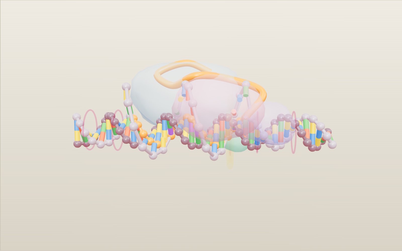
How CRISPR-Cas9 gene editing works
A guide RNA reads just 20 letters of DNA, and that is usually enough to pick out one spot among the 3 billion base pairs of a human genome.
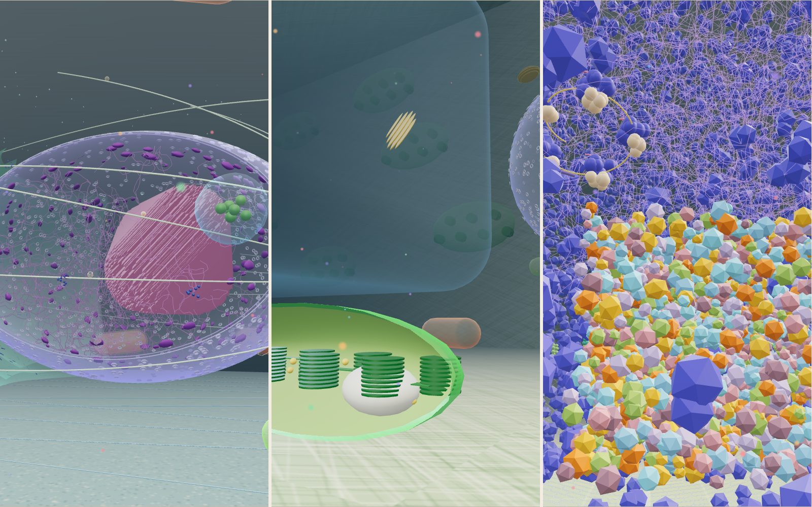
Walk inside a cell: animal, plant and bacterium
Grown a million times, a mitochondrion is about the size of a sofa, a whole E. coli is 2 m long, and the kinesin towing a vesicle past you walks at 0.8 metres per second.
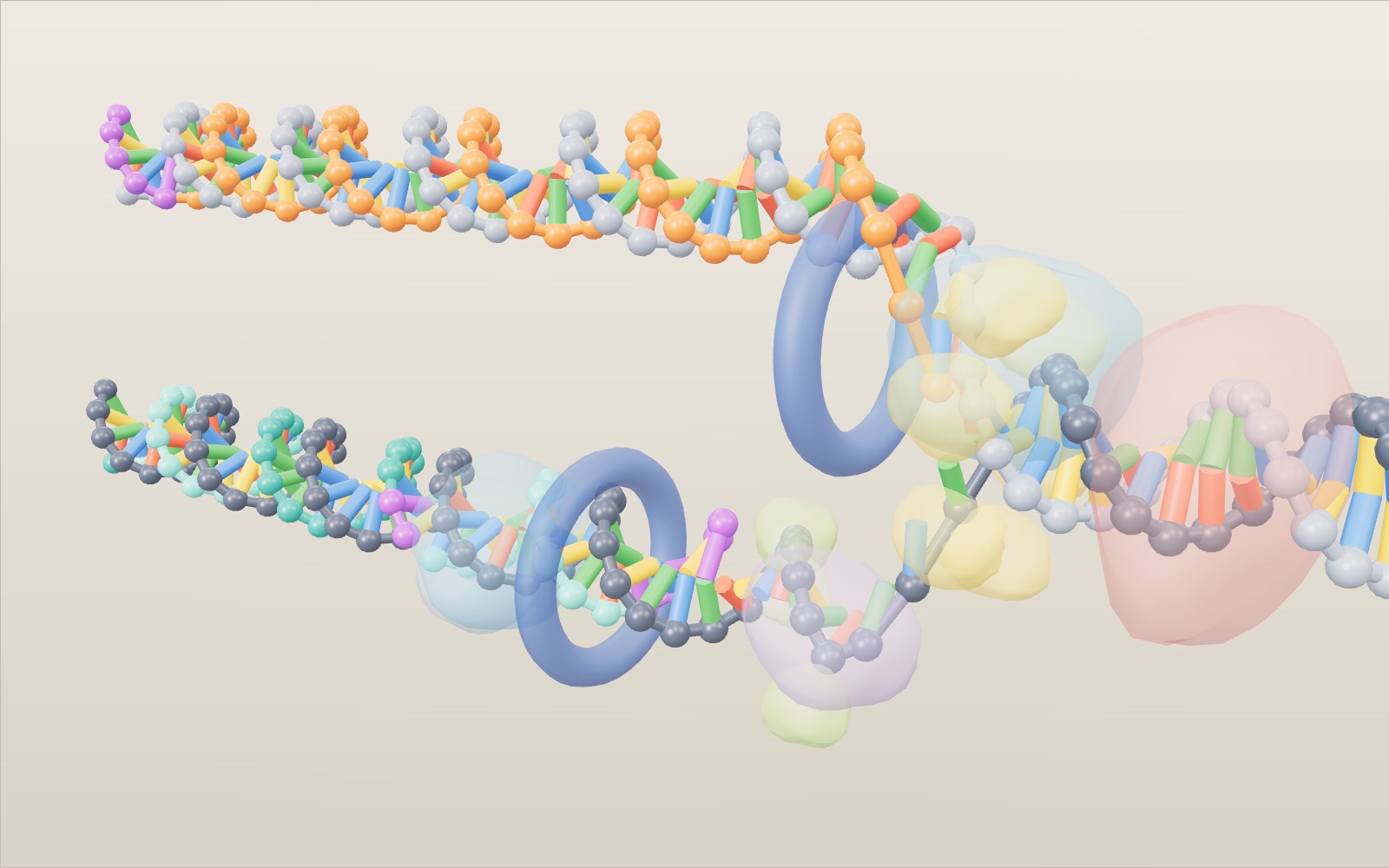
How DNA replication works, step by step: Okazaki fragments in 3D
A DNA polymerase can only add to one end of a strand, so at every replication fork one new strand grows in a single piece and the other in short fragments.
How nerve signals travel
A nerve cell sits at about −70 mV, and when a stimulus lifts it to about −55 mV it fires a spike that is always the same size.
Giant sequoia forest: walk under the largest trees
Stand at the foot of the General Sherman Tree, 83.8 m tall and about 2,200 years old, then climb its trunk and see how hard the water up there is pulled.
Dinosaur size comparison to humans: 172 life-size animals
A 600 m park of 172 prehistoric animals at their real size, from the first dinosaurs to T. rex, laid out by age.
How photosynthesis works
A leaf in full sun pulls carbon dioxide out of air that holds only about 426 parts per million of it, and builds sugar from it.
Coral bleaching causes: why warmer water turns reefs white
Corals get most of their food from algae living in their tissue, and when the sea stays more than 1 °C above its usual summer high for weeks, they lose those algae and turn white.
How do bats see with sound? Echolocation explained
An echo from a moth 1 m away comes back to a bat in under 6 thousandths of a second, and that delay is how the bat knows the distance.
What happens to your body in space without a suit
Without a suit you would have about 10 seconds of useful consciousness, and if air came back within a minute or so you might well recover. You would not explode.
Fly with life-size pterosaurs
Quetzalcoatlus spanned about 10 m, close to the 11 m wing of a Cessna 172, and standing it was as tall as a giraffe. Watch it pass over a Cretaceous cliff, then fly with 35 pterosaurs from three ages.
Giants of the sea: megalodon and the blue whale
Swim beside 48 sea giants at their real size, from a 28 m blue whale to megalodon, Livyatan and Mosasaurus, in eight seas from today back to the Triassic.