FIG. 54Walk-throughs
Walk inside a cell: animal, plant and bacterium
Grown a million times, a mitochondrion is about the size of a sofa, a whole E. coli is 2 m long, and the kinesin towing a vesicle past you walks at 0.8 metres per second.
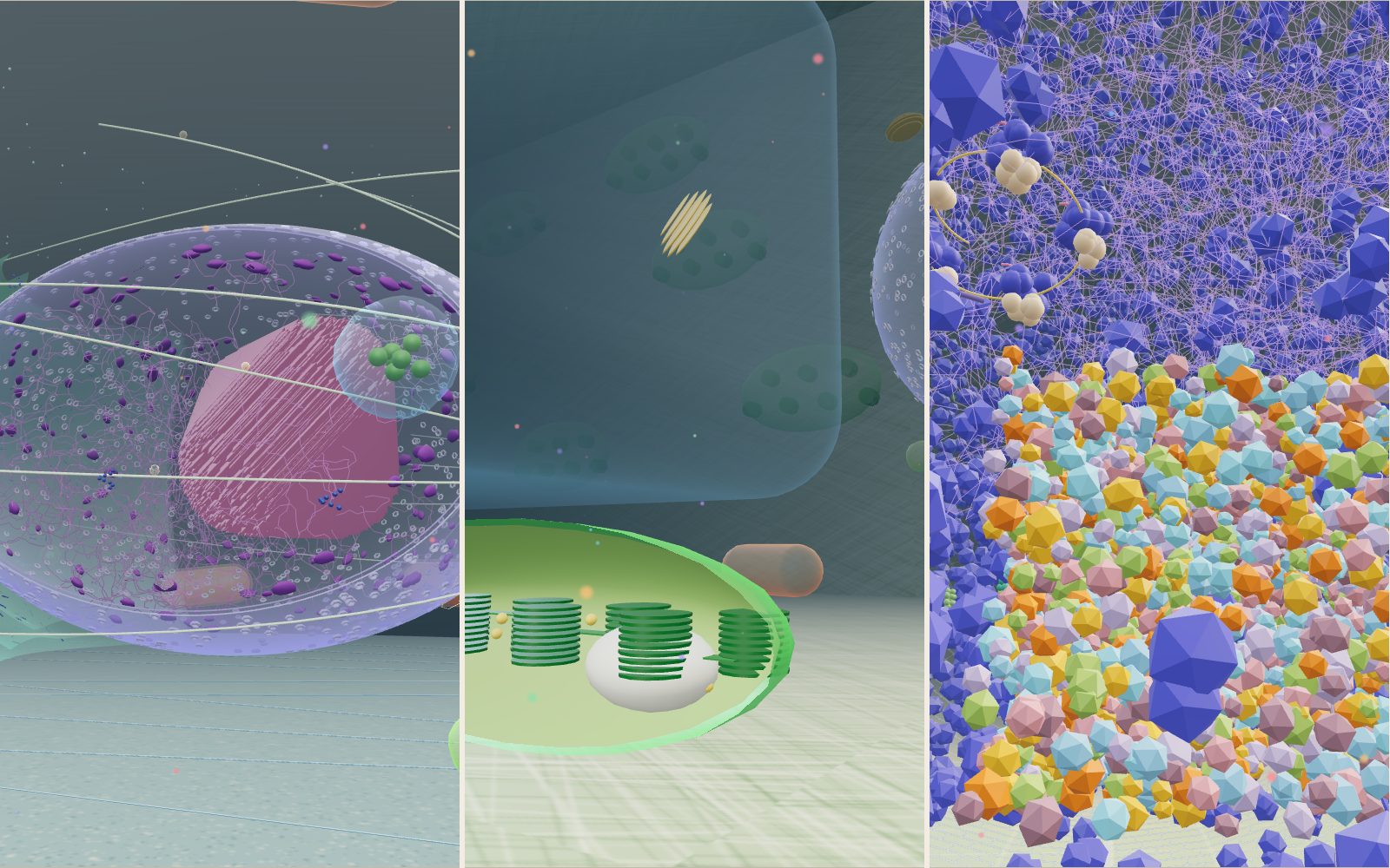
This device cannot show the 3D model. You can read the guide steps below as text.
1 / 14 A cell a million times largerYou are standing on the bottom membrane of a cell grown a million times, so your eyes are 1.6 µm above it. Move with WASD, the arrow keys or the joystick, and rise or sink with R and F or the arrows on the right. The nucleus ahead has a window cut into it. The Guide book button at the top right lists 78 things to visit.
Drag to turn. Scroll or pinch to zoom.
What you are standing in
A cell is too small to picture. The usual drawing in a school book puts the nucleus, a few mitochondria and some wavy lines of ER inside a circle, all about the same size and all floating in empty space. This exhibit takes one animal cell, enlarges it a million times and lets you walk around inside it at an ordinary eye height. Everything is drawn to the sizes measured in real cells, so a mitochondrion is a capsule you could sit on and a ribosome is a dark bead the size of a large marble.
The cell is shaped like one spread out on a glass dish: a low dome 30 µm long, 24 µm wide and 8 µm tall in the middle. You start on its floor, which is the part of the plasma membrane stuck to the glass. The nucleus fills the middle, with rough ER wrapped around its left side and the Golgi stack on its right. Mitochondria and lysosomes are scattered around, microtubules fan out from a point beside the nucleus, and small vesicles ride along them towards the edge of the cell.
Why the model is a million times larger
The obvious choice would be to make the cell large enough that your body fits inside it. A magnification of 100,000 is not enough. At that scale a 20 µm cell becomes a 2 m bubble, and the nucleus would be smaller than your head. A person 1.6 m tall would stand 16 µm high, taller than the whole cell.
At a magnification of 1,000,000 the numbers become easy to read. One metre in the model is one micrometre in the cell, and one millimetre is one nanometre. Your eye height of 1.6 m stands for 1.6 µm. The scale box in the top left corner shows this, and its bar is redrawn for whatever is straight ahead of you. If you look at a wall 3 m away, the bar shows how long 1 µm looks at that distance.
Sizes in the model
These are the main parts and the sizes they are built to. The real figures are ranges, because cells differ from one type to the next.
| Part | Real size | Size in the model |
|---|---|---|
| Whole cell (spread on glass) | 20 to 40 µm across | 30 × 24 × 8 m |
| Nucleus | 2 to 10 µm across | 9 × 6 × 7.6 m |
| Mitochondrion | about 1 µm wide, 2 µm long | 0.84 to 1 m wide, 2 to 2.8 m long |
| Lysosome | 0.1 to 1.2 µm | 0.5 to 0.7 m |
| Nuclear pore | about 120 nm | 12 cm |
| Microtubule | 25 nm wide | 2.5 cm wide |
| Ribosome | 25 to 30 nm | about 2.8 cm |
| Smooth ER tube | 30 to 100 nm | 6 cm |
| Plasma membrane | 4 to 10 nm thick | 4 to 10 mm thick |
| Kinesin step | 8 nm | 8 mm |
The cell's volume works out at about 3,000 µm³. Cell Biology by the Numbers uses 2,000 µm³ as a rule of thumb for a human cell, with most falling between 500 and 4,000 µm³, and it uses 3,000 µm³ for a characteristic mammalian cell in its worked examples. HeLa cells spread on a slide reach about 40 µm across while staying only a few micrometres tall, which is why the model is a low dome rather than a ball.
Checking a few numbers yourself
Stand next to the cut-open mitochondrion near the start. It is 1 m wide, so its top is a little below your eye level. In the real cell that is 1 µm, about a hundredth of the width of a human hair.
The membrane is the hardest part to see. At 4 to 10 nm thick it is 4 to 10 mm in the model, about as thick as a pencil, wrapped around a room 30 m long. Cell Biology by the Numbers compares it to the skin of an aircraft against the width of its body. That is why one patch on the right-hand wall is drawn 20 times thicker than the rest. In that patch each lipid head is 16 mm across instead of 0.8 mm, and you can count the two layers.
The DNA in the nucleus is the number that does not fit at all. A human cell carries about 2 m of DNA (two copies of a genome of about 3 billion base pairs, 0.3 nm per base pair). In the model that would be 2,000 km of thread packed into a ball 9 m long. The purple threads you see inside the window are a small sample drawn to show the texture.
The membrane in cross-section
A phospholipid has a head that mixes with water and two tails that do not. In water the tails hide from it by lining up tail to tail, which gives two layers with heads on both outer faces. Each layer is about 2 nm deep, so the core of the bilayer is about 4 nm thick. Proteins sit in this film. The blue barrel in the magnified patch is a channel that spans both layers, and the protein with green beads on the outside carries a chain of sugars.
The membrane is fluid. Lipids drift sideways past their neighbours all the time, but they very rarely flip from one layer to the other, so the two layers can keep different mixes of lipids. The model holds them still.
The nucleus and its pores
The nuclear envelope is two membranes, an inner and an outer, with a gap of a few tens of nanometres between them. The window cut into the front of the nucleus shows both rims, about 4 cm apart in the model. The outer membrane carries on into the rough ER, which is why the ER sheets in the model start right next to the nucleus.
Nuclear pores pass through both membranes at once. Each is a ring of proteins about 120 nm across with a channel through the middle, and a vertebrate nucleus has about 2,000 of them. Messenger RNA and ribosome subunits go out through the pores, and proteins needed inside the nucleus come in. The model shows 300 to 1,100 pores depending on your device.
Inside, the nucleolus is a dense region with no membrane around it, drawn 2 µm across in the model. It forms around the genes for ribosomal RNA, and the two halves of the ribosome are put together there before they are exported.
Where proteins are made and packed
A ribosome reads messenger RNA and joins amino acids into a chain. Ribosomes floating in the cytosol make proteins that stay in the cytosol. When the start of a new protein carries a signal for the ER, the ribosome docks onto the ER and feeds the growing chain through the membrane into the sac, which is what makes the ER look rough. Smooth ER has no ribosomes. It is a network of tubes 30 to 100 nm wide that makes lipids and stores calcium.
From the ER, proteins travel in small vesicles to the Golgi apparatus. A mammalian cell has 40 to 100 Golgi stacks, usually gathered near the centrosome, and each stack has four to eight cisternae. The model shows one stack of six. Proteins enter on the cis side, nearest the ER, have sugars trimmed and added as they move through, and leave from the trans side in vesicles headed for the membrane, the lysosomes or other compartments.
Kinesin walking on a microtubule
Microtubules are hollow tubes about 25 nm wide, usually built from 13 rows of tubulin. Each row is a chain of alpha and beta tubulin pairs that repeats every 8 nm. Most microtubules start at the centrosome beside the nucleus and run out towards the edge of the cell. They are stiff and nearly straight over the distances inside a cell.
Kinesin-1 has two motor heads joined by a long coiled stalk, and a tail that holds the cargo. It walks hand over hand, as PDB-101 describes it. One head stays bound to the microtubule while the other lets go, swings forward past it and binds 16 nm further on. Each step moves the cargo 8 nm and uses one ATP. Cell Biology by the Numbers gives about 800 nm per second for conventional kinesin in the test tube, with about 100 steps before it falls off.
At a million times life size, 800 nm per second becomes 0.8 m per second, a slow walking pace. But the heads take about 100 steps a second, far too fast to follow on screen. The guide step about kinesin therefore switches on slow motion at 1/20 of real speed, so you can see each head swing past the other.
Mitochondria and lysosomes
Mitochondria have two membranes. The outer one is smooth. The inner one is folded into cristae, which give it a large surface for the proteins that make ATP. Cell Biology by the Numbers describes a typical mitochondrion as a capsule roughly 2 µm long and 1 µm across, though in many cells they join into branching networks tens of micrometres long. The model uses ten separate capsules and cuts one open so you can see the cristae.
Lysosomes are bags of more than 60 kinds of digestive enzyme, and a cell normally has hundreds of them. The model shows six. Their inside is kept at pH 4.5 to 5, and the enzymes work best at that acidity. The cytosol around them is at about pH 7.2, where the enzymes do not work properly, so a leaking lysosome does not digest the rest of the cell.
Shrinking ten and a hundred times
At a million times life size the cell is easy to walk through, but anything smaller than about 20 nm is a crumb at your feet. A ribosome is the size of a marble and a potassium channel is 8 mm across. To see molecules you need a second step of magnification, and the Size buttons in the top right give you two more: ×10 and ×100.
The buttons do not rebuild the cell. They shrink you. At ×10 the scale is 10,000,000 to 1, one metre in front of you is 100 nm, and your eyes are 160 nm above the floor. At ×100 the scale is 100,000,000 to 1, one metre is 10 nm, and you stand 16 nm tall, about the height of a proteasome. The box in the top left corner always shows the current magnification, what one metre stands for, and your real height, so the numbers stay honest whichever step you are on. The cell itself does not change. A mitochondrion that was the size of a sofa becomes a building 100 m long, and walking past it at ×100 would take minutes.
Molecules at their true size
Molecules are drawn as bead models, with one bead for each folded part of a protein, in the style of the illustrations on RCSB PDB-101. They are built at their true size and placed where they work, then hidden until you are close enough for them to show. From across the cell they cost nothing to draw.
There are four places to find them in the animal cell:
- On the floor membrane, near the front right. Potassium channels, sodium-potassium pumps, receptors and aquaporins sit in a patch of membrane paved with lipid heads 0.8 nm apart. A clathrin-coated pit, a cage about 100 nm wide, is pulling a bud of membrane inwards. Actin filaments 7 nm wide run across the floor, with a myosin V motor walking on one of them, and a rope-like intermediate filament lies nearby.
- In the crowd beside them. A box 100 nm on each side is filled with proteins at the density of real cytosol, about 3 million per cubic micrometre. At ×100 it is a wall of molecules you can push into. A proteasome, an Hsp90 chaperone, a glycogen granule and a string of ribosomes reading one messenger RNA sit at its edge.
- In the nucleus. A stretch of DNA wound round nucleosomes, with RNA polymerase II making a strand of RNA, and a nuclear pore complex in full detail on the envelope.
- On organelles. A ribosome docked on the rough ER through its Sec61 channel, a row of ATP synthases at the cut edge of the open mitochondrion, and a dynein motor under the kinesin track.
The densities are real where the guide book says so (the crowd box, the lipid floor) and thinned out elsewhere, because a real membrane is so packed with protein that the channels would be hard to pick out.
A plant cell at the same scale
The Cell button switches to a leaf cell built to the same scale. It is 40 µm long, 24 µm wide and 20 µm tall, inside a wall of cellulose. Cell Biology by the Numbers describes plant cells of this kind with most of their volume taken up by a vacuole, and that is the first thing you notice: a translucent block fills the middle, and the rest of the cell is squeezed into a thin layer around it and a room at one end where the nucleus sits.
Chloroplasts lie on the floor under the vacuole and against one wall. They are drawn 5 µm long, inside the range of 4 to 6 µm given for vascular plants, and one is cut open to show its grana, stacks of flat thylakoid sacs, and a grain of starch. On the far wall a pit field of plasmodesmata, channels about 50 nm wide, connects this cell to its neighbour. Along the near wall, organelles stream round a loop of actin cables at 4 µm per second, inside the range of 1 to 100 µm per second measured for cytoplasmic streaming. At ×100 inside the cut chloroplast you can find Rubisco, the enzyme that fixes carbon dioxide, and photosystem II, which splits water and releases oxygen.
A bacterium at the same scale
The third cell is E. coli. Cell Biology by the Numbers uses about 1 µm wide, 2 µm long and 1 µm³ in volume as the rule of thumb, so at a million times life size the whole organism is a capsule 2 m long lying on the dish, the size of one of the animal cell's mitochondria. That is not a coincidence of the model. Mitochondria descend from bacteria that were taken in by another cell in the distant past, and they still keep a small genome of their own. The grid on the dish is drawn in 1 µm squares.
At ×1 you are too big to go inside, so the walk starts at ×10. Inside, ribosomes fill most of the space around the nucleoid, the tangle of the chromosome in the middle with no membrane around it. The envelope overhead has three layers: the inner membrane, a thin net of peptidoglycan and the outer membrane. Flagellar motors about 45 nm across sit in the envelope, each turning a corkscrew filament about 100 times a second, and a lattice of chemoreceptors at one end tells the motors when to reverse. Because there is no nucleus in the way, ribosomes start reading messenger RNA while it is still being made, which the model shows at two places near the start.
The guide book
The Guide book button opens a list of 78 things to visit: 49 in the animal cell, 14 in the plant cell and 15 in the bacterium. You can search by name, filter by cell and by kind (organelles, molecules and processes) and sort by size. Choosing one switches cell and size if needed and walks you to a spot in front of it. A card then shows its real size, how many a cell has, a short description, its sources, and a bar comparing its size with a protein, a ribosome, E. coli, an animal cell and your own height at the current size.
Seven processes, the stages of mitosis, the cell cycle, the cell plate of a dividing plant cell and binary fission in bacteria, are not played out in the 3D cells. Their cards show a small drawing instead.
What a million times does to speeds and distances
Because 1 m in the model is 1 µm in the cell, every length can be converted by moving the unit up by six steps. A cell 20 µm across is 20 m across, about the length of a bus and a half laid end to end. A ribosome 25 nm across is 25 mm, 2.5 cm, the size of a large marble. A protein of 5 nm is 5 mm, a grain of rice seen end on. A membrane 5 nm thick is 5 mm thick, a layer you could hold between two fingers around a hall 30 m long.
Speeds scale the same way, because time is not stretched. Kinesin walks at about 800 nm per second, which is 0.8 m per second in the model. A protein that drifts a few micrometres in a second covers a few metres. You walk at about 1.4 m per second, which is 1.4 µm per second in the real cell, a little faster than a kinesin. Crossing the 30 m length of the cell takes you about 21 seconds. At ×100 the same walk is 14 nm per second, and the Walk control has a fast setting for that reason.
The units hide one trap. Areas scale with the square and volumes with the cube. A mitochondrion of 1 µm³ is 1 m³ in the model, a cube a metre on each side. The whole cell, about 3,000 µm³, is 3,000 m³, a hall larger than an Olympic swimming pool, which holds about 2,500 m³. The numbers in the table above are lengths. When this guide gives a count per cell, it means per 3,000 of those cubic metres.
How many molecules, and how long they take to cross
Cell Biology by the Numbers gives about 3 million proteins in every cubic micrometre of a cell, so a human cell of 2,000 to 4,000 µm³ holds on the order of 10 billion (10¹⁰) proteins. For the 3,000 µm³ of this model that works out at 3,000 × 3 million, about 9 billion. In the model, one cubic metre of space around you holds 3 million of them. The average gap between neighbouring proteins is the cube root of 1 µm³ divided by 3 million, about 0.007 µm or 7 nm, which is 7 mm on screen. A typical protein is about 5 nm across, so the crowd is nearly touching. This is the density the crowd box reproduces. Everything else in the model is much emptier than that.
Molecules in a cell do not travel in straight lines. They are knocked about by water molecules, a motion called diffusion, and the average time to cover a distance x grows with the square of that distance. For a particle moving in one dimension it is t ≈ x² / 2D, where D is the diffusion constant. In three dimensions the 2 becomes a 6. The book gives D as about 100 µm²/s for a protein of average size in water and about 10 µm²/s inside a cell. Putting in the numbers:
- Across an E. coli cell, x = 1 µm: t ≈ 1 / 60, about 0.02 s. The book quotes roughly 0.01 s.
- Across a HeLa cell, x = 20 µm: t ≈ 400 / 60, about 7 s. The book quotes about 10 s.
- Along an axon 1 cm long, x = 10,000 µm: t ≈ 10⁸ / 60, about 1.7 million s, roughly three weeks. The book says about two weeks, since it rounds D and the factor.
A motor moving at 1 µm per second would take 2 to 3 hours to cover 1 cm, which is why nerve cells carry cargo on microtubules rather than waiting for it to diffuse. At the scale of a cell, though, the two methods are close. At 20 µm a motor needs 20 s and diffusion needs under 10 s, so a small cell can let molecules diffuse and keeps motors for cargo that is large or must go to a particular place.
Common misunderstandings
- "Organelles float far apart in empty water." In a real cell the cytosol is a crowded gel and the organelles are packed close, with the ER stretching almost everywhere. The open room you walk through in the model is a simplification so that you can move.
- "Molecules know where to go." Most reach their partner by chance, by diffusion, and react only when they happen to meet in the right orientation. Motors like kinesin are the exception, since they follow tracks, but the cargo still has to find the track.
- "A kinesin walks like a person." It looks like it from the animation, but the heads are small enough to be pushed about by the surrounding water all the time. The walk is a ratchet in which each ATP makes one head swing forward and bind, so the jiggling is turned into steps in one direction.
- "The nucleus is the cell's brain, so it controls everything directly." The nucleus stores and copies the genome and makes RNA, but proteins are made in the cytosol and on the ER, and many of the cell's decisions are made by signals arriving at the membrane. Mitochondria even keep a small genome of their own.
- "All cells look like the drawing in the textbook." The model shows one cell type spread on glass, with the sizes of the ones commonly studied. A red blood cell has no nucleus and a muscle fibre has many, and cell volumes range over several orders of magnitude.
How the model differs from a real cell
- The cytosol is nearly empty here. A real cell holds about 3 million proteins in each cubic micrometre. The floating specks around you are a few thousand at most, and they drift slowly instead of jostling at high speed.
- Ribosomes are thinned out. The model draws a few thousand. A real cell has far more, packed so densely on the rough ER that the sheets look studded.
- Actin and intermediate filaments are sparse. Bundles of actin cross the floor and a few intermediate filaments wrap the nucleus, but the dense actin mesh under the membrane is left out so the paths stay clear.
- Molecules are hidden from afar. The molecule models appear only when you are close enough to see them, and they sit still instead of tumbling.
- Most organelles keep still. In a living cell, mitochondria move, split and join, and the ER changes shape from minute to minute.
- The nucleus has a window. A real envelope is closed apart from its pores.
- Colours are invented. Organelles have no colour of their own at this size. Electron microscopes show them in grey, and fluorescence images use dyes.
Moving around
On a computer, use WASD or the arrow keys to walk and drag with the mouse to look around. R lifts you up and F lets you sink back down. Shift makes you move faster. Clicking the floor walks you there. On a phone, the left half of the screen is a joystick and the right half turns your view. The two arrows on the right lift you up or down.
The panel on the right of the stage has three rows. Cell switches between the animal cell, the plant cell and the bacterium. Size shrinks you to ×10 or ×100 and back. Walk sets your pace to slow, normal or fast, which helps at ×100, where normal walking covers only 13 nm a second. The small map in the top left shows where you are from above; click it to walk to that spot.
Name tags appear when you come close to something, and at ×100 they label single molecules. The All name tags control shows more of them at a time. The Parts list and the Guide book both take you straight to whatever you choose.
Questions
Is this what a real cell looks like inside?
The sizes and positions follow real measurements, but a real cell is far more crowded. Proteins, RNA and small molecules fill almost every gap, and the organelles are packed closer than in the model. If it were drawn at its true density you would not be able to see more than a few micrometres in any direction.
Why does the cell have a floor?
Most animal cells studied in the lab are grown on glass or plastic, where they spread out flat and stick to the surface. The floor is the part of the plasma membrane touching the dish. In a tissue the cell would be surrounded by other cells on all sides.
How fast does kinesin really move?
Conventional kinesin moves about 800 nm per second in the test tube and can go faster inside cells. That is 8 nm steps taken about 100 times a second. At the model's scale this is 0.8 m per second, so without slow motion it walks past you at a stroll while its heads blur.
Can a vesicle really ride a microtubule this far?
Yes. Kinesins carry vesicles, organelles and other cargo along microtubules, and dynein carries cargo the other way, towards the centrosome. A single kinesin usually lets go after about a hundred steps, but cargo often carries several motors, so it can be passed along and keep moving.
Why are mitochondria shown as separate capsules?
That is the textbook picture and it is true for many cells, especially in electron microscope sections. In living cells they often form long branching networks that keep splitting and fusing. Separate capsules are easier to see.
How many ribosomes does a cell have?
It depends on the kind of cell and how fast it is growing. Cell Biology by the Numbers shows that a fast-growing E. coli has about 72,000 ribosomes, more than a third of its dry mass. An animal cell is a thousand or more times larger by volume and has far more again, so the few thousand in this model are a thin sample.
Why can I float inside the nucleus?
The window is a cut-away, like those used in museum models of engines. In a real cell the envelope is closed and nothing as large as you could pass through a nuclear pore.
Why can't I walk into the bacterium at the normal size?
Because at a million times life size it is only 2 m long and 1 m wide, and you are 1.6 m tall with your eyes. Shrinking ten times makes you 160 nm tall and the bacterium a tunnel 10 m wide. This is the same size relationship as in real life: an E. coli cell is about as large as one mitochondrion, and an animal cell could hold thousands of them.
Are the molecules really packed this tightly?
In the crowd box and the lipid floor, yes. Cell Biology by the Numbers puts the protein content of cells at about 3 million proteins per cubic micrometre, and the box is filled at that density. Elsewhere the molecule models are thinned out so you can find single ones, and the specks floating around you are only a token amount.
Why is the plant cell so empty in the middle?
The middle is the central vacuole, a sac of water, salts and pigments. In many leaf cells it takes up 80% or more of the volume. Its pressure against the wall is what keeps soft parts of a plant stiff, and a plant wilts when the vacuoles lose water.
How do the size buttons change what I see?
They make you smaller while the cell stays the same. At ×10 every length in front of you is ten times larger than before, so a ribosome that was a marble becomes a football. The scale box always shows the current magnification, what one metre stands for, and your own height in real units.
Terms
- Organelle: a part of the cell with its own job, such as the nucleus or a mitochondrion.
- Cytosol: the watery gel that fills the cell around the organelles.
- Phospholipid bilayer: the two layers of lipid molecules that make up cell membranes.
- Nuclear envelope: the double membrane around the nucleus.
- Nuclear pore complex: a protein ring about 120 nm wide that forms a channel through the nuclear envelope.
- Chromatin: DNA wound around histone proteins, the form DNA takes in the nucleus.
- Endoplasmic reticulum (ER): a network of membrane sacs and tubes. Rough ER carries ribosomes; smooth ER does not.
- Cisterna: one flattened sac of the ER or the Golgi apparatus.
- Cristae: the folds of the inner mitochondrial membrane.
- Microtubule: a hollow tube of tubulin about 25 nm wide.
- Motor protein: a protein that uses ATP to move along a filament. Kinesin and dynein walk on microtubules.
- ATP: adenosine triphosphate, the small molecule cells use to carry energy.
- Vacuole: the large sac of water in a plant cell, wrapped in a membrane called the tonoplast.
- Chloroplast: the organelle where photosynthesis happens, with stacks of thylakoids inside.
- Plasmodesma: a channel through the plant cell wall that joins two neighbouring cells.
- Nucleoid: the region of a bacterium where its chromosome lies, with no membrane around it.
- Peptidoglycan: the net of sugar chains and short peptides that forms a bacterial cell wall.
- Flagellar motor: the rotary motor in a bacterial envelope that turns the flagellum.
- Magnification: how many times larger the model is than the real thing. ×10 and ×100 multiply the starting 1,000,000.
Sources
- Milo, R. and Phillips, R., Cell Biology by the Numbers: How big is a human cell? https://book.bionumbers.org/how-big-is-a-human-cell/ (checked 16 October 2026)
- Milo, R. and Phillips, R., Cell Biology by the Numbers: How big are nuclei? https://book.bionumbers.org/how-big-are-nuclei/ (checked 16 October 2026)
- Milo, R. and Phillips, R., Cell Biology by the Numbers: How big are mitochondria? https://book.bionumbers.org/how-big-are-mitochondria/ (checked 16 October 2026)
- Milo, R. and Phillips, R., Cell Biology by the Numbers: How big is the endoplasmic reticulum of cells? https://book.bionumbers.org/how-big-is-the-endoplasmic-reticulum-of-cells/ (checked 16 October 2026)
- Milo, R. and Phillips, R., Cell Biology by the Numbers: What is the thickness of the cell membrane? https://book.bionumbers.org/what-is-the-thickness-of-the-cell-membrane/ (checked 16 October 2026)
- Milo, R. and Phillips, R., Cell Biology by the Numbers: What are the sizes of the cell's filaments? https://book.bionumbers.org/what-are-the-sizes-of-the-cells-filaments/ (checked 16 October 2026)
- Milo, R. and Phillips, R., Cell Biology by the Numbers: How fast do molecular motors move on cytoskeletal filaments? https://book.bionumbers.org/how-fast-do-molecular-motors-move-on-cytoskeletal-filaments/ (checked 16 October 2026)
- Milo, R. and Phillips, R., Cell Biology by the Numbers: How big are the molecular machines of the central dogma? https://book.bionumbers.org/how-big-are-the-molecular-machines-of-the-central-dogma/ (checked 16 October 2026)
- Milo, R. and Phillips, R., Cell Biology by the Numbers: The geography of the cell. https://book.bionumbers.org/the-geography-of-the-cell/ (checked 16 October 2026)
- Milo, R. and Phillips, R., Cell Biology by the Numbers: How big are genomes? https://book.bionumbers.org/how-big-are-genomes/ (checked 16 October 2026)
- Milo, R. and Phillips, R., Cell Biology by the Numbers: How many ribosomes are in a cell? https://book.bionumbers.org/how-many-ribosomes-are-in-a-cell/ (checked 16 October 2026)
- RCSB PDB-101, Molecule of the Month: Kinesin. https://pdb101.rcsb.org/motm/64 (checked 16 October 2026)
- RCSB PDB-101, Molecule of the Month: Nuclear Pore Complex. https://pdb101.rcsb.org/motm/205 (checked 16 October 2026)
- RCSB PDB-101, Molecule of the Month: Ribosomal Subunits. https://pdb101.rcsb.org/motm/10 (checked 16 October 2026)
- Nature Education, Scitable: Mitochondria. https://www.nature.com/scitable/topicpage/mitochondria-14053590/ (checked 16 October 2026)
- Milo, R. and Phillips, R., Cell Biology by the Numbers: How large are chloroplasts?. https://book.bionumbers.org/how-large-are-chloroplasts/ (checked 17 October 2026)
- Milo, R. and Phillips, R., Cell Biology by the Numbers: How big is an E. coli cell and what is its mass?. https://book.bionumbers.org/how-big-is-an-e-coli-cell-and-what-is-its-mass/ (checked 17 October 2026)
- Milo, R. and Phillips, R., Cell Biology by the Numbers: What is the frequency of rotary molecular motors?. https://book.bionumbers.org/what-is-the-frequency-of-rotary-molecular-motors/ (checked 17 October 2026)
- Milo, R. and Phillips, R., Cell Biology by the Numbers: How fast do proteasomes degrade proteins?. https://book.bionumbers.org/how-fast-do-proteasomes-degrade-proteins/ (checked 17 October 2026)
- Milo, R. and Phillips, R., Cell Biology by the Numbers: How many ions pass through an ion channel per second?. https://book.bionumbers.org/how-many-ions-pass-through-an-ion-channel-per-second/ (checked 17 October 2026)
- Milo, R. and Phillips, R., Cell Biology by the Numbers: What are the rates of membrane transporters?. https://book.bionumbers.org/what-are-the-rates-of-membrane-transporters/ (checked 17 October 2026)
- Milo, R. and Phillips, R., Cell Biology by the Numbers: How long do the different stages of the cell cycle take?. https://book.bionumbers.org/how-long-do-the-different-stages-of-the-cell-cycle-take/ (checked 17 October 2026)
- Milo, R. and Phillips, R., Cell Biology by the Numbers: How many proteins are in a cell?. https://book.bionumbers.org/how-many-proteins-are-in-a-cell/ (checked 17 October 2026)
- Milo, R. and Phillips, R., Cell Biology by the Numbers: How big is the average protein?. https://book.bionumbers.org/how-big-is-the-average-protein/ (checked 17 October 2026)
- Milo, R. and Phillips, R., Cell Biology by the Numbers: What is faster, transcription or translation?. https://book.bionumbers.org/what-is-faster-transcription-or-translation/ (checked 17 October 2026)
- Milo, R. and Phillips, R., Cell Biology by the Numbers: What is the pH of a cell?. https://book.bionumbers.org/what-is-the-ph-of-a-cell/ (checked 17 October 2026)
- Milo, R. and Phillips, R., Cell Biology by the Numbers: What is the range of cell sizes and shapes?. https://book.bionumbers.org/what-is-the-range-of-cell-sizes-and-shapes/ (checked 17 October 2026)
- OpenStax, Biology 2e, 4.3 Eukaryotic Cells. https://openstax.org/books/biology-2e/pages/4-3-eukaryotic-cells (checked 17 October 2026)
- OpenStax, Biology 2e, 4.2 Prokaryotic Cells. https://openstax.org/books/biology-2e/pages/4-2-prokaryotic-cells (checked 17 October 2026)
- OpenStax, Biology 2e, 4.4 The Endomembrane System and Proteins. https://openstax.org/books/biology-2e/pages/4-4-the-endomembrane-system-and-proteins (checked 17 October 2026)
- OpenStax, Biology 2e, 4.5 The Cytoskeleton. https://openstax.org/books/biology-2e/pages/4-5-the-cytoskeleton (checked 17 October 2026)
- OpenStax, Biology 2e, 4.6 Connections between Cells and Cellular Activities. https://openstax.org/books/biology-2e/pages/4-6-connections-between-cells-and-cellular-activities (checked 17 October 2026)
- OpenStax, Biology 2e, 10.2 The Cell Cycle. https://openstax.org/books/biology-2e/pages/10-2-the-cell-cycle (checked 17 October 2026)
- OpenStax, Biology 2e, 10.5 Prokaryotic Cell Division. https://openstax.org/books/biology-2e/pages/10-5-prokaryotic-cell-division (checked 17 October 2026)
- OpenStax, Biology 2e, 15.5 Ribosomes and Protein Synthesis. https://openstax.org/books/biology-2e/pages/15-5-ribosomes-and-protein-synthesis (checked 17 October 2026)
- OpenStax, Biology 2e, 15.2 Prokaryotic Transcription. https://openstax.org/books/biology-2e/pages/15-2-prokaryotic-transcription (checked 17 October 2026)
- OpenStax, Biology 2e, 3.4 Proteins. https://openstax.org/books/biology-2e/pages/3-4-proteins (checked 17 October 2026)
- RCSB PDB-101, Molecule of the Month: ATP Synthase. https://pdb101.rcsb.org/motm/72 (checked 17 October 2026)
- RCSB PDB-101, Molecule of the Month: Potassium Channels. https://pdb101.rcsb.org/motm/38 (checked 17 October 2026)
- RCSB PDB-101, Molecule of the Month: Sodium-Potassium Pump. https://pdb101.rcsb.org/motm/118 (checked 17 October 2026)
- RCSB PDB-101, Molecule of the Month: Adrenergic Receptors. https://pdb101.rcsb.org/motm/100 (checked 17 October 2026)
- RCSB PDB-101, Molecule of the Month: Aquaporin. https://pdb101.rcsb.org/motm/173 (checked 17 October 2026)
- RCSB PDB-101, Molecule of the Month: Clathrin. https://pdb101.rcsb.org/motm/88 (checked 17 October 2026)
- RCSB PDB-101, Molecule of the Month: Proteasome. https://pdb101.rcsb.org/motm/166 (checked 17 October 2026)
- RCSB PDB-101, Molecule of the Month: Hsp90. https://pdb101.rcsb.org/motm/108 (checked 17 October 2026)
- RCSB PDB-101, Molecule of the Month: Chaperones. https://pdb101.rcsb.org/motm/32 (checked 17 October 2026)
- RCSB PDB-101, Molecule of the Month: RNA Polymerase. https://pdb101.rcsb.org/motm/40 (checked 17 October 2026)
- RCSB PDB-101, Molecule of the Month: Nucleosome. https://pdb101.rcsb.org/motm/7 (checked 17 October 2026)
- RCSB PDB-101, Molecule of the Month: Ribosome. https://pdb101.rcsb.org/motm/121 (checked 17 October 2026)
- RCSB PDB-101, Molecule of the Month: Elongation Factors. https://pdb101.rcsb.org/motm/81 (checked 17 October 2026)
- RCSB PDB-101, Molecule of the Month: Actin. https://pdb101.rcsb.org/motm/19 (checked 17 October 2026)
- RCSB PDB-101, Molecule of the Month: Microtubules. https://pdb101.rcsb.org/motm/175 (checked 17 October 2026)
- RCSB PDB-101, Molecule of the Month: Dynein. https://pdb101.rcsb.org/motm/176 (checked 17 October 2026)
- RCSB PDB-101, Molecule of the Month: Myosin. https://pdb101.rcsb.org/motm/18 (checked 17 October 2026)
- RCSB PDB-101, Molecule of the Month: Rubisco. https://pdb101.rcsb.org/motm/11 (checked 17 October 2026)
- RCSB PDB-101, Molecule of the Month: Photosystem II. https://pdb101.rcsb.org/motm/59 (checked 17 October 2026)
- RCSB PDB-101, Molecule of the Month: Flagellar Motor. https://pdb101.rcsb.org/motm/300 (checked 17 October 2026)
- RCSB PDB-101, Molecule of the Month: Pilus Machine. https://pdb101.rcsb.org/motm/211 (checked 17 October 2026)
- RCSB PDB-101, Molecule of the Month: Penicillin-binding Proteins. https://pdb101.rcsb.org/motm/29 (checked 17 October 2026)
- RCSB PDB-101, Molecule of the Month: Catalase. https://pdb101.rcsb.org/motm/57 (checked 17 October 2026)
- RCSB PDB-101, Molecule of the Month: SNARE Proteins. https://pdb101.rcsb.org/motm/167 (checked 17 October 2026)
- RCSB PDB-101, Molecule of the Month: Vacuolar ATPase. https://pdb101.rcsb.org/motm/219 (checked 17 October 2026)
- RCSB PDB-101, Molecule of the Month: Glycogen Phosphorylase. https://pdb101.rcsb.org/motm/24 (checked 17 October 2026)
- RCSB PDB-101, Molecule of the Month: Cellulose Synthase. https://pdb101.rcsb.org/motm/254 (checked 17 October 2026)
- BioNumbers BNID 106729: Diameter of peroxisome (0.1 to 1.0 µm). https://bionumbers.hms.harvard.edu/bionumber.aspx?id=106729 (checked 17 October 2026)
- BioNumbers BNID 113930: Organelle numbers in COS-7 cells (lipid droplets, peroxisomes, lysosomes about 90 to 190 each). https://bionumbers.hms.harvard.edu/bionumber.aspx?id=113930 (checked 17 October 2026)
- BioNumbers BNID 104378: Diameter of late endosome in HeLa cells (0.76 µm). https://bionumbers.hms.harvard.edu/bionumber.aspx?id=104378 (checked 17 October 2026)
- BioNumbers BNID 104387: Diameter of late endosome, Rab9-depleted HeLa cells (0.42 µm). https://bionumbers.hms.harvard.edu/bionumber.aspx?id=104387 (checked 17 October 2026)
- Fujimoto and Parton, Not Just Fat: The Structure and Function of the Lipid Droplet, Cold Spring Harb Perspect Biol 2011 (PMC3039932). https://pmc.ncbi.nlm.nih.gov/articles/PMC3039932/ (checked 17 October 2026)
- BioNumbers BNID 105741: Size of canonical centriole (about 0.5 × 0.2 µm). https://bionumbers.hms.harvard.edu/bionumber.aspx?id=105741 (checked 17 October 2026)
- BioNumbers BNID 106125: Diameter of intermediate filaments (8 to 10 nm). https://bionumbers.hms.harvard.edu/bionumber.aspx?id=106125 (checked 17 October 2026)
- BioNumbers BNID 112144: Number of clathrin-coated structures per cell (about 9,000). https://bionumbers.hms.harvard.edu/bionumber.aspx?id=112144 (checked 17 October 2026)
- BioNumbers BNID 112147: Clathrin-coated pit mean lifetime (about 40 s). https://bionumbers.hms.harvard.edu/bionumber.aspx?id=112147 (checked 17 October 2026)
- BioNumbers BNID 104385: Diameter of endocytic vesicle (100 ± 30 nm). https://bionumbers.hms.harvard.edu/bionumber.aspx?id=104385 (checked 17 October 2026)
- MetaCyc (SRI International): glycogen, β particles about 20 to 30 nm. https://biocyc.org/META/new-image?object=Glycogens (checked 17 October 2026)
- BioNumbers BNID 107396: Beta hairpin folding time (6 µs). https://bionumbers.hms.harvard.edu/bionumber.aspx?id=107396 (checked 17 October 2026)
- BioNumbers BNID 111269: GFP folding time (600 s). https://bionumbers.hms.harvard.edu/bionumber.aspx?id=111269 (checked 17 October 2026)
- BioNumbers BNID 102727: Diameter of a vesicle (40 to 80 nm). https://bionumbers.hms.harvard.edu/bionumber.aspx?id=102727 (checked 17 October 2026)
- OpenStax Biology 2e, 5.4 Bulk Transport. https://openstax.org/books/biology-2e/pages/5-4-bulk-transport (checked 17 October 2026)
- BioNumbers BNID 102983: Diameter of entire mitotic chromosome (1,400 nm). https://bionumbers.hms.harvard.edu/bionumber.aspx?id=102983 (checked 17 October 2026)
- Betterton and McIntosh, Regulation of chromosome speeds in mitosis, Cell Mol Bioeng 2013 (PMC4578309). https://pmc.ncbi.nlm.nih.gov/articles/PMC4578309/ (checked 17 October 2026)
- Focus on cell walls, Plant Physiology 2024 (PMC10756750). https://pmc.ncbi.nlm.nih.gov/articles/PMC10756750/ (checked 17 October 2026)
- BioNumbers BNID 100594: Plasmodesmata diameter (50 nm, 20 to 200). https://bionumbers.hms.harvard.edu/bionumber.aspx?id=100594 (checked 17 October 2026)
- BioNumbers BNID 100593: Plasmodesmata per µm² of cell wall (2 to 10). https://bionumbers.hms.harvard.edu/bionumber.aspx?id=100593 (checked 17 October 2026)
- Zeeman et al., Starch Synthesis in Arabidopsis. Granule Synthesis, Composition, and Structure, Plant Physiol 2002 (PMC161669). https://pmc.ncbi.nlm.nih.gov/articles/PMC161669/ (checked 17 October 2026)
- Goldstein and van de Meent, A physical perspective on cytoplasmic streaming, Interface Focus 2015 (PMC4590424). https://pmc.ncbi.nlm.nih.gov/articles/PMC4590424/ (checked 17 October 2026)
- BioNumbers BNID 104947: Width of cell envelope of E. coli B (28.7 nm). https://bionumbers.hms.harvard.edu/bionumber.aspx?id=104947 (checked 17 October 2026)
- BioNumbers BNID 108581: Thickness of peptidoglycan (Gram-negative a few nm). https://bionumbers.hms.harvard.edu/bionumber.aspx?id=108581 (checked 17 October 2026)
- BioNumbers BNID 109343: Diameter of bacterial flagellum (about 20 nm). https://bionumbers.hms.harvard.edu/bionumber.aspx?id=109343 (checked 17 October 2026)
- BioNumbers BNID 100095: Typical length of E. coli flagella (5 to 10 µm). https://bionumbers.hms.harvard.edu/bionumber.aspx?id=100095 (checked 17 October 2026)
- BioNumbers BNID 100105: Diameter of E. coli pili (7 nm). https://bionumbers.hms.harvard.edu/bionumber.aspx?id=100105 (checked 17 October 2026)
- BioNumbers BNID 100104: Length of E. coli pili (1 to 2 µm). https://bionumbers.hms.harvard.edu/bionumber.aspx?id=100104 (checked 17 October 2026)
- BioNumbers BNID 101473: Number of pili per E. coli cell (100 to 300). https://bionumbers.hms.harvard.edu/bionumber.aspx?id=101473 (checked 17 October 2026)
- Briegel et al., Universal architecture of bacterial chemoreceptor arrays, PNAS 2009 (PMC2761316; E. coli array about 53,000 nm², about 5,200 receptors). https://pmc.ncbi.nlm.nih.gov/articles/PMC2761316/ (checked 17 October 2026)
- OpenStax Microbiology, 3.3 Unique Characteristics of Prokaryotic Cells. https://openstax.org/books/microbiology/pages/3-3-unique-characteristics-of-prokaryotic-cells (checked 17 October 2026)
- RCSB PDB 3DKN: Sec61 in the canine ribosome-channel complex. https://www.rcsb.org/structure/3dkn (checked 17 October 2026)
- BioNumbers BNID 100121: Diameter of 70S ribosome (26 nm). https://bionumbers.hms.harvard.edu/bionumber.aspx?id=100121 (checked 17 October 2026)
- BioNumbers BNID 100483: Ribosome diameter (30 nm). https://bionumbers.hms.harvard.edu/bionumber.aspx?id=100483 (checked 17 October 2026)
More exhibits
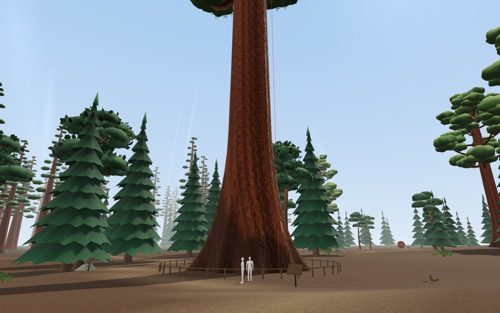
Giant sequoia forest: walk under the largest trees
Stand at the foot of the General Sherman Tree, 83.8 m tall and about 2,200 years old, then climb its trunk and see how hard the water up there is pulled.
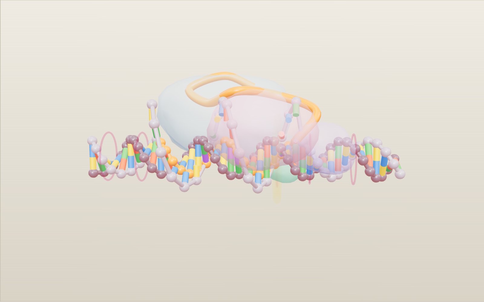
How CRISPR-Cas9 gene editing works
A guide RNA reads just 20 letters of DNA, and that is usually enough to pick out one spot among the 3 billion base pairs of a human genome.
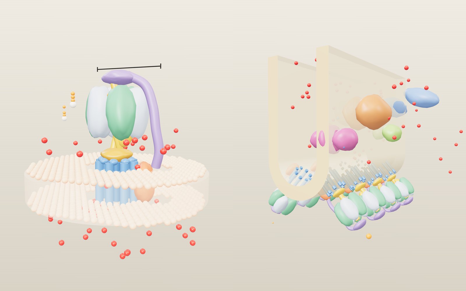
How ATP synthase works
In mitochondria the rotor of ATP synthase turns about 100 times a second, and every full turn releases three molecules of ATP.
Dinosaur size comparison to humans: 172 life-size animals
A 600 m park of 172 prehistoric animals at their real size, from the first dinosaurs to T. rex, laid out by age.
How DNA replication works, step by step: Okazaki fragments in 3D
A DNA polymerase can only add to one end of a strand, so at every replication fork one new strand grows in a single piece and the other in short fragments.
Walk among ice age animals
A 600 m park of 65 animals from the last ice age at their real size, from woolly mammoths to sabre-toothed cats, on a time slider that runs 50,000 years.