Loading the model
This device cannot show the 3D model. You can read the guide steps below as text.
1 / 14 A cell a million times larger You are standing on the bottom membrane of a cell grown a million times, so your eyes are 1.6 µm above it. Move with WASD, the arrow keys or the joystick, and rise or sink with R and F or the arrows on the right. The nucleus ahead has a window cut into it. The Guide book button at the top right lists 78 things to visit.
Drag to turn. Scroll or pinch to zoom.
Guide Parts Controls
01 A cell a million times larger You are standing on the bottom membrane of a cell grown a million times, so your eyes are 1.6 µm above it. Move with WASD, the arrow keys or the joystick, and rise or sink with R and F or the arrows on the right. The nucleus ahead has a window cut into it. The Guide book button at the top right lists 78 things to visit.
02 The membrane, 20 times thicker The real membrane is only 4 to 10 nm thick, which is 4 to 10 mm at this scale. Here it is drawn 20 times thicker so you can see the two layers of lipids. Their heads face the water on either side and their oily tails meet in the middle.
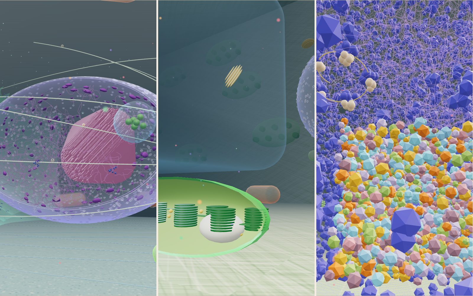
03 Pores in the nuclear envelope The nucleus is wrapped in two membranes about 40 nm apart, and the window shows both of them cut through. The rings dotted across the surface are nuclear pores, each about 120 nm wide. A vertebrate nucleus has around 2,000 of them.
04 Inside the nucleus You have floated in through the window. The purple threads are chromatin, which is DNA wound around proteins, and the darker lumps near the envelope are tightly packed stretches of it. The large knot ahead is the nucleolus, where ribosomes are assembled.
05 Rough ER and its ribosomes These flattened sacs carry on from the outer membrane of the nucleus. The dark dots on them are ribosomes, 25 to 30 nm across, feeding new proteins into the sac as they grow. Further to the left, the tubes without ribosomes are smooth ER.
06 The Golgi stack Six curved sacs sit stacked like plates. Proteins arrive from the ER on the side facing the nucleus and are trimmed and labelled as they pass through. Small vesicles bud off the rims on the far side and carry them away.
07 Kinesin on a microtubule This is slow motion at 1/20 of real speed. The red motor is kinesin. Its two heads take turns stepping 8 nm along one of the 13 rows of tubulin, using one ATP per step, while the long stalk tows a vesicle. At real speed it covers about 0.8 µm a second.
08 A mitochondrion cut open Mitochondria in this cell are about 1 µm wide and 2 to 3 µm long. This one is cut open, so you can see the folds of its inner membrane, called cristae, where most of the cell's ATP is made. The pink ball to the right is a lysosome.
09 100 times smaller: proteins in the membrane You have shrunk 100 times, so the scale is now 100,000,000 to 1 and you are 16 nm tall. The floor is the membrane, paved with lipid heads 0.8 nm apart. The blue rings are potassium channels, the green blobs are sodium-potassium pumps and the purple bundles are receptors. The cage further back is a clathrin-coated pit about 100 nm wide.
10 The crowd in the cytosol This box of proteins is drawn at the real density, about 3 million proteins in every cubic micrometre. At its edge sit a proteasome, the barrel that cuts up old proteins, an Hsp90 chaperone and a row of ribosomes reading one messenger RNA.
11 A plant cell This is a leaf cell at the same scale as the animal cell, 40 µm long inside a wall of cellulose. The large translucent block is the central vacuole, which fills most of the cell and pushes everything else into a thin layer and a room at this end. Watch the side wall: organelles stream along cables of actin at about 4 µm a second.
12 Inside a chloroplast One chloroplast is cut open. The green stacks are grana, piles of flat thylakoid sacs about 0.5 µm across where light is captured, and the white lump is a grain of starch. Shrink 100 times and walk in to see Rubisco and photosystem II.
13 A bacterium is 2 m long here At the same million-fold scale an E. coli cell is a capsule 2 m long and 1 m wide, about as big as a mitochondrion. The orange corkscrews are its flagella, 20 nm thick and several micrometres long. You are too big to fit inside.
14 Inside E. coli You are now ten times smaller, 160 nm tall. The dark beads are ribosomes, tens of thousands of them, and the purple tangle in the middle is the chromosome, with no nuclear envelope around it. The three layers of the envelope curve overhead.
Cell membrane A double layer of lipids 4 to 10 nm thick, which is 4 to 10 mm at this scale. One patch on the right-hand wall is drawn 20 times thicker so the two layers show. Nucleus 9 µm long and 6 µm tall here, inside a double membrane dotted with pores about 120 nm wide. A window is cut into the front so you can float inside. Nucleolus A dense knot about 2 µm across inside the nucleus. The RNA and proteins of ribosomes are put together here before they leave through the pores. Rough endoplasmic reticulum Flattened sacs wrapped around the left side of the nucleus and joined to its outer membrane. The ribosomes on them make proteins for membranes and for export. Smooth endoplasmic reticulum A network of tubes 30 to 100 nm wide with no ribosomes. It makes lipids and holds calcium for the cell to release when signalled. Ribosomes Protein-making machines 25 to 30 nm across, about the size of a large marble here. Some sit on the rough ER and some float free in small clusters. Golgi apparatus A stack of six curved sacs about 2.5 µm across. Proteins enter on the side facing the nucleus and leave from the far side in vesicles. Vesicles Membrane bubbles about 80 nm across. They ride the microtubules out to the membrane, where they fuse and empty their cargo. Microtubules and kinesin Hollow tubes 25 nm wide that fan out from the centrosome. Kinesin walks along one of them towing a vesicle, 8 nm per step. Mitochondria Ten capsules about 1 µm wide and 2 to 3 µm long. The one near the front is cut open to show the folds of its inner membrane. Lysosomes Six bags of digestive enzymes, 0.5 to 0.7 µm across. Their inside is kept at pH 4.5 to 5, far more acidic than the cytosol around them. Plant cell A leaf cell 40 µm long inside its wall, with a central vacuole, chloroplasts (one cut open), plasmodesmata and cytoplasm streaming around the vacuole. Bacterium (E. coli) A cell 2 µm long, which is only 2 m at this scale. You start inside it after shrinking ten times, among ribosomes, the nucleoid and the flagellar motors. Pause