FIG. 59How It Works
How DNA replication works, step by step: Okazaki fragments in 3D
A DNA polymerase can only add to one end of a strand, so at every replication fork one new strand grows in a single piece and the other in short fragments.
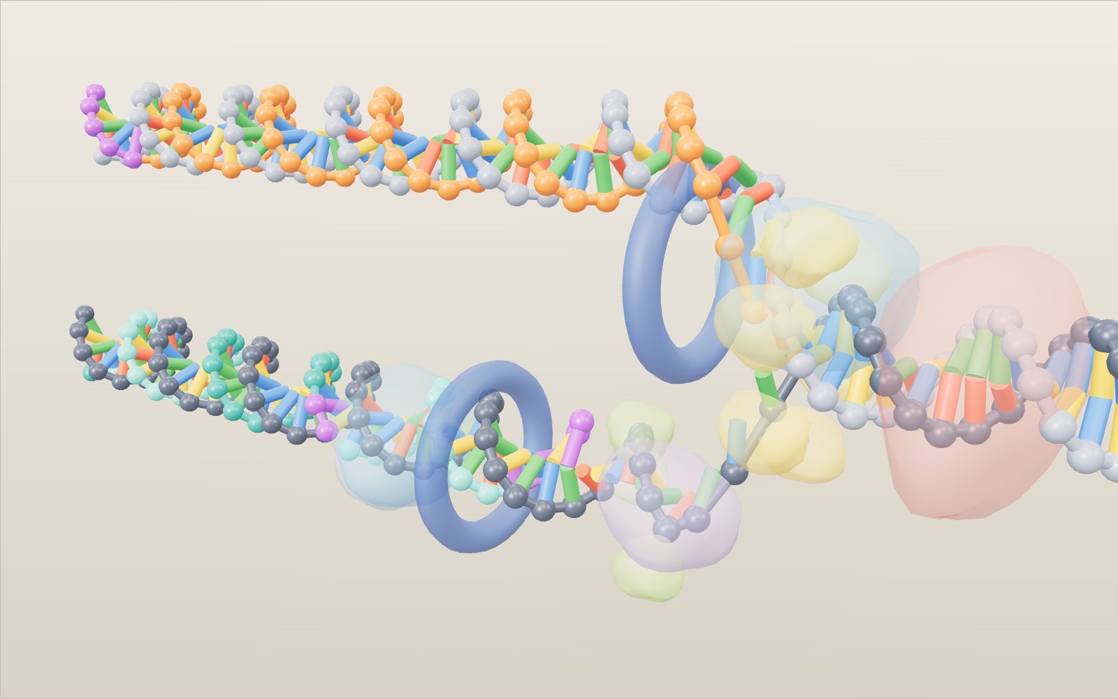
This device cannot show the 3D model. You can read the guide steps below as text.
1 / 11 84 base pairs of DNA, to scaleThis stretch is about 29 nm long. The bars below it give the scale: one turn is 10.5 base pairs (3.57 nm) in solution, while textbooks round it to 10 base pairs and 3.4 nm. The sequence is made up for the model.
Drag to turn. Scroll or pinch to zoom.
How does a cell copy its DNA before it divides?
DNA replication works in a few steps at a moving Y-shaped fork. Helicase unzips the double helix and primase lays a short RNA primer. DNA polymerase can add nucleotides only to a 3′ end, so the leading strand grows in one piece while the lagging strand is made backwards in Okazaki fragments, and DNA polymerase I and ligase then swap the primers for DNA and seal the gaps. A human fork copies 50 to 100 nucleotides per second, and a human cell copies its 6.4 billion base pairs in an S phase of about 8 to 10 hours.
A cut on your hand heals because skin cells divide, and each new cell needs its own full set of DNA. In a human cell that means copying about 6 billion base pairs before the cell splits in two. The copying is done by a group of proteins working at a moving Y-shaped junction, the replication fork. They have to unwind the helix, start each new strand, build it base by base, catch their own mistakes and join the pieces they leave behind.
This exhibit builds one fork at molecular scale. A stretch of 84 base pairs lies across the stage. A helicase opens it, primase lays short RNA starters, two DNA polymerases build the new strands, DNA polymerase I and DNA ligase tidy up the lagging strand, and at the end two double helices move apart. The opening shot starts right beside the helicase as it unzips the helix. Drag the yellow helicase ring to pull the fork yourself, press Pair the next base to put in bases as the polymerase does, and tap any enzyme to read its job. Use the slider to scrub through the process, the Speed control to change the pace and with it the length of the Okazaki fragments, and the Proofreading switch to see what happens when the polymerase stops checking its work. The facts come from the OpenStax Biology textbook, Nature Education, the US National Human Genome Research Institute and the original 1958 paper by Meselson and Stahl. The sources are listed at the end.
The double helix at the scale of the model
DNA is two strands wound round each other. Each strand is a chain of sugars and phosphates, and each sugar carries one of four bases: A, C, G or T. A on one strand pairs with T on the other, and C pairs with G. That rule is what makes copying possible: once the strands are apart, each one carries all the information needed to rebuild its partner. In the model the bases are coloured A green, T red, G yellow and C blue. RNA uses U in place of T, and U is purple.
Neighbouring base pairs are 0.34 nm apart and the helix is 2 nm wide. Textbooks often say the helix turns once every 10 base pairs, or 3.4 nm. That figure comes from X-ray pictures of DNA fibres. Measured in solution, as it is in a cell, DNA turns a little less tightly: James Wang found 10.4 base pairs per turn in 1979, and 10.5 is the value most often used. The model uses 10.5, so one turn is 10.5 × 0.34 = 3.57 nm. The two bars under the model show both: one model turn, and the textbook 3.4 nm for comparison. The whole model is 84 × 0.34 = 28.6 nm long.
The two strands run in opposite directions. Chemists number the carbon atoms of each sugar, and the ends of a strand are called 5′ and 3′ after them. A strand that runs 5′ to 3′ from left to right is paired with one that runs 5′ to 3′ from right to left. This detail, antiparallel strands, decides most of what happens at the fork.
What opens the helix and keeps it open
The enzyme that opens DNA is helicase. It breaks the hydrogen bonds between the paired bases and uses energy from ATP to do it. Where it works, the double helix splits into two single strands, and the shape that forms is the replication fork. In the model the helicase is a yellow ring sitting at the point where the strands part, and it moves along as the fork advances.
Pulling two twisted strands apart has a side effect. The DNA in front of the fork gets wound tighter, the way a twisted rope kinks up when you pull its strands apart at one end. OpenStax describes how topoisomerase prevents this overwinding by making temporary nicks in the helix ahead of the fork and resealing them. In the model it is the pink blob on the unopened DNA, a few turns ahead of the helicase.
The opened single strands would pair up again if they were left alone. Single-strand binding proteins coat them and keep them apart until a polymerase arrives. In the model they are the small green blobs. Watch where they gather: almost all of them sit on the bottom template, because that strand waits longest before it is copied. The reason is the next part of the story.
Why a polymerase needs a primer
DNA polymerase adds nucleotides one at a time, each one matched to the base opposite it on the template. The energy for each new bond comes from the incoming nucleotide itself, which arrives as a triphosphate. But the enzyme has two strict limits. It can only add to the 3′ end of a strand, and it needs a strand to be there already. It cannot start from nothing.
Primase solves the starting problem. It is an RNA polymerase, and RNA polymerases can start a new chain without a partner. Primase writes a short piece of RNA, about 5 to 10 nucleotides long according to OpenStax, complementary to the template. That primer gives DNA polymerase a free 3′ end to extend. In the model primers are 5 nucleotides long and drawn as purple beads. One primer at the far left starts the top new strand. The bottom new strand needs a fresh primer again and again.
One direction only, and what it does to the two strands
Because polymerase can only add to a 3′ end, every new strand grows in the 5′ to 3′ direction. Now look at the two templates. On the top template, the new strand's 5′ to 3′ direction points towards the fork. The polymerase can follow the helicase and keep adding to the same strand as more template is exposed. This continuous strand is the leading strand, orange in the model, and it is built from a single primer.
On the bottom template the new strand has to run the other way, away from the fork. As the fork opens, fresh template appears behind the polymerase rather than in front of it. The only way to copy it is to start a new piece near the fork and build backwards until the piece meets the one made before. Each of these pieces is an Okazaki fragment, and the strand built from them is the lagging strand, teal in the model, with alternate fragments in two shades.
Switch off Base pair colours in the Controls tab to see this clearly. Old strands turn grey and the two kinds of new strand keep their own colours. The leading strand is one long orange line. The lagging strand is a row of short teal pieces with purple primers at their right-hand ends.
Okazaki fragments on the lagging strand
The lagging strand repeats one cycle. Primase lays a primer close to the fork. The lagging-strand polymerase, held on the DNA by a ring-shaped sliding clamp, extends that primer away from the fork until it runs into the previous fragment. Then it lets go and moves to the next primer. In the model you can watch the second polymerase and its dark blue clamp jump back towards the fork after finishing each piece.
Real fragments are much longer than the model's 14 nucleotides. A review by Balakrishnan and Bambara gives about 1,200 nucleotides for Okazaki fragments in bacteria and only about 200 in eukaryotic cells, where the length follows the spacing of nucleosomes. The model shortens them so several fit on the screen at once. The Okazaki row in the readout counts two numbers: how many fragments have been started and how many have been joined to their neighbour.
Swapping primers for DNA and sealing the nicks
A finished fragment still has RNA at one end, and a new strand of DNA should not contain RNA. In bacteria, DNA polymerase I removes each primer with its exonuclease activity and fills the gap with DNA, using the DNA of the fragment behind it as its starting point. In eukaryotic cells the job is done differently: polymerase δ displaces the primer into a flap, and flap endonuclease 1 (FEN1) cuts it away. RNase H can also contribute to primer removal, but RNase H and FEN1 are different enzymes. The model shows the bacterial version, a pale blue enzyme that turns the purple beads into teal ones.
After the swap, the two pieces of DNA lie end to end but are not yet joined. One phosphodiester bond is missing, a gap called a nick. DNA ligase forms that bond between the 3′ end of one piece and the 5′ end of the next. In the model the orange ligase sits on the nick for a moment, a ring flashes, and the backbone link appears. That is when the joined count goes up.
How the polymerase takes back a wrong base
Polymerases sometimes put in the wrong base. Nature Education gives the rate as about 1 in every 100,000 nucleotides. A wrong base sits badly in the helix, and its 3′ end is in the wrong position for the next addition. The polymerase notices, backs up, cuts the wrong nucleotide out with its 3′ to 5′ exonuclease activity and puts in the right one. This is proofreading, and OpenStax describes it as the polymerase reading each newly added base before adding the next one.
The model has two planted mistakes on the leading strand, a T opposite a G and a G opposite a T. Both are wobble pairs, the kind of mispairing Nature Education describes. With Proofreading on, the polymerase stops at each one: the wrong base turns magenta, swings out, disappears and is replaced. While it is stopped, the helicase waits too, so the fork falls behind. Switch Proofreading off and play the same stretch. The fork reaches each point sooner and the Okazaki counts run ahead, but the two wrong bases stay in the new strand, ringed in magenta, and the readout says how many were left in.
Checking the numbers
The model's default pace is 5 base pairs per second. A human replication fork moves at 50 to 100 nucleotides per second according to OpenStax, and Nature Education gives about 40. So the model runs about 10 to 20 times slower than a real human fork. A bacterial fork is much faster: E. coli copies its 4.6 million base pair chromosome in about 42 minutes with two forks, which works out to roughly 1,000 nucleotides per second at each fork.
Can one human fork copy a whole genome? At 50 nucleotides per second, 6 billion base pairs would take 6,000,000,000 ÷ 50 = 120 million seconds, almost four years. Cells avoid that by starting many forks at once; OpenStax says humans can have up to 100,000 origins of replication. The National Human Genome Research Institute puts the copying time for one human cell at several hours.
Errors add up in the same way. Copying 6 billion base pairs means laying down about 12 billion new nucleotides, one opposite each template base. At 1 mistake in 100,000, that is about 120,000 wrong bases per division before any checking. Proofreading fixes about 99 percent of them, which leaves roughly 1,200. Mismatch repair after replication brings the number lower still. In the model, 2 planted mistakes among 84 base pairs is wildly more than real life; they are there so you can see the correction happen.
What the model leaves out
The model shows one fork. Real replication starts at an origin and opens a bubble with two forks moving apart, and the two forks in E. coli travel in opposite directions around its circular chromosome and meet, each copying approximately half the circle. The model's fragments and primers are shortened, and only two mistakes are planted, at fixed places.
The proteins are soft blobs, not measured shapes, and they are see-through so you can follow the DNA. Their placement follows textbook diagrams: topoisomerase ahead of the fork, single-strand binding proteins on bare template, a clamp ring around each new double helix next to its polymerase. The two polymerases are drawn as separate blobs so each new strand is easy to follow. The leading-strand primer at the far left is also left in place, because the fork that would remove it is off the left edge.
The protein names follow bacteria (DNA polymerase III does the main copying, DNA polymerase I swaps primers). Eukaryotic cells use different enzymes for the same steps: pol ε on the leading strand, pol δ on the lagging strand, PCNA as the clamp and FEN1 with contributions from RNase H in primer processing. They also have to remove and replace histones as the fork passes, which OpenStax suggests is part of the reason eukaryotic forks are slower.
Several numbers on the stage are model values. The fragment length is the Speed times a priming interval of 2.8 seconds of screen time, kept between 7 and 36 nucleotides, so 5 bp/s gives 14 nt. Pairing a base by hand moves the fork one base pair and skips the two programmed mistakes, so only your own picks are wrong. The calculators below the stage assume evenly spaced origins that all fire together and forks that never stall, and they scale the cell turnover of a 70 kg adult in proportion to body weight.
How we know each new helix keeps one old strand
When James Watson and Francis Crick published the double helix in 1953, they noted that the pairing rule suggested a copying mechanism. There were three ideas for how it might work. In semiconservative replication each new helix keeps one old strand and gains one new strand. In conservative replication the old helix stays whole and a completely new one is built beside it. In dispersive replication old and new DNA are mixed in pieces along both strands.
Matthew Meselson and Franklin Stahl, at the California Institute of Technology, tested them in 1958. They grew E. coli for many generations on a medium whose only nitrogen was the heavy isotope nitrogen-15, then moved the cells to ordinary nitrogen-14 and took samples as they divided. DNA spun in a caesium chloride gradient settles where its density matches the solution, so heavy and light DNA form separate bands. Before the switch there was one heavy band. After one generation there was a single band halfway between heavy and light, which ruled out conservative replication. After two generations there were two bands, one hybrid and one light, which ruled out the dispersive model. Each new helix had kept one old strand. Switch on Meselson and Stahl in the Controls tab to see these three tubes in the readout.
The fragments of the lagging strand were found by Reiji Okazaki and his colleagues, who reported in 1968 that newly made DNA first appears as short pieces that are later joined into long strands. The enzymes themselves were studied in the test tube earlier. Arthur Kornberg and Severo Ochoa shared the 1959 Nobel Prize in Physiology or Medicine "for their discovery of the mechanisms in the biological synthesis of ribonucleic acid and deoxyribonucleic acid", in the words of the Nobel committee.
How many forks would finish a human genome in hours?
A fork speed is a distance along a template per unit time, not the combined output of every polymerase in a cell. At an assumed constant 1,000 nt/s, two bacterial forks cover 2,000 base pairs of parental chromosome per second. A 4,600,000 bp chromosome therefore needs 4,600,000 ÷ 2,000 = 2,300 seconds, or about 38 minutes. The textbook's roughly 42 minutes is compatible with these rounded inputs; the calculation is not a prediction of the time between bacterial cell divisions. Under favourable growth conditions, bacteria can begin another round of replication before the previous one finishes.
For a human example, use a rounded haploid genome of 3,200,000,000 bp and assume that 10,000 evenly spaced origins all fire together. Each origin sends out two forks at 50 nt/s, giving 20,000 forks and a total coverage rate of 1,000,000 bp/s. Dividing genome length by that rate gives 3,200 seconds, about 53 minutes. There are on average 320,000 bp between origins, and each fork travels half that distance before meeting its neighbour. The same answer follows from 160,000 ÷ 50.
This deliberately ideal calculation is shorter than the several hours needed by a real human cell. Origins do not all fire together; different chromosome regions replicate at different times, and forks encounter chromatin and other obstacles. If only 2,000 origins were active together under the same assumptions, the calculation would give 16,000 seconds, about 4.4 hours. These origin counts are assumed examples, not measurements of a particular tissue. The comparison explains why counting potential origins alone cannot determine the length of S phase.
Most human body cells have two chromosome sets. At the same rounded scale that means 6,400,000,000 bp to duplicate, rather than the haploid 3,200,000,000 bp. Doubling both the DNA amount and the number of active origins leaves the ideal completion time unchanged. The earlier 6 billion figure is a coarser diploid approximation, not a different rule for counting base pairs.
From an incorrect nucleotide to a lasting mutation
A wrong insertion, a mismatch that escapes proofreading, and a mutation retained after repair are different counts. The review by Fijalkowska and colleagues gives a useful bacterial example: initial insertion errors around 10⁻⁵, an escape fraction around 10⁻² after proofreading, and around 10⁻³ after mismatch repair. Multiplying gives 10⁻¹⁰ errors per copied base per round. These are approximate contributions, dependent on the mismatch and sequence, not universal constants for every polymerase.
For an arithmetic exercise, retain the earlier rounded diploid 6 billion bp genome. Copying both parental strands incorporates about 12 billion new nucleotides. At 10⁻⁵, that gives 120,000 initial errors; at 10⁻⁷ after proofreading it gives 1,200. A final rate of 10⁻⁹ would leave an expected 12 errors across the replication products, while 10⁻¹⁰ would give 1.2. A fractional expectation describes an average over many divisions; a cell cannot contain a fraction of a changed base. Proofreading alone does not provide the whole reduction from 10⁻⁵ to 10⁻⁹ or 10⁻¹⁰.
Do not turn those illustrative totals into a claim that every human division creates exactly that many mutations. A mismatch still contains conflicting instructions on its two strands. Repair can remove it, or a later round of copying can establish the altered sequence in a descendant. The products also separate into daughter cells. DNA damage, cell type and the fidelity of different enzymes affect observed mutation counts. The screen's fixed pair of errors tests the correction sequence, not a mutation-rate distribution; replaying it does not sample another cell.
How many fragments must be finished?
Fragment length changes the workload without changing the amount of DNA that must be copied. Using the review's approximate 1,200 nt bacterial fragment length, the lagging-strand contribution for a 4,600,000 bp circular chromosome is of order 4,600,000 ÷ 1,200, or about 3,800 fragments. Why not divide twice the chromosome length? At each fork one of the two newly made strands is lagging. Summed over both forks, the length made discontinuously is approximately one chromosome length, not both new strands together.
With a rounded diploid human genome of 6,400,000,000 bp and fragments averaging 200 nt, the corresponding estimate is 32,000,000 fragments. End effects and differences between regions prevent these divisions from being exact counts. Even so, they show why primer removal and ligation cannot be rare finishing touches. A cell has to process millions of junctions during a single S phase. Short fragments in eukaryotes are associated with nucleosome organisation, rather than meaning that human polymerases can synthesize only 200 nucleotides before losing all activity.
The exhibit compresses each fragment to 14 nt and each primer to 5 nt. That makes a primer occupy a much larger fraction of a fragment than in the biological examples. Do not estimate the amount of RNA in a chromosome from the proportion of purple beads on screen. Watch instead for the order of events: the primer supplies the start, DNA replaces the primer, and ligase closes the remaining backbone nick. Ligase does not read a template and fill a long missing stretch of DNA by itself.
Chromosome ends and PCR solve different problems
The ordinary lagging-strand cycle depends on a neighbouring fragment supplying an extendable 3′ end when a primer is removed. At the end of a linear chromosome, that arrangement runs out. Telomeres are repeated DNA with associated protective proteins at chromosome ends. In cells with active telomerase, the enzyme uses its own RNA as a template to extend a DNA end; other replication machinery can then copy complementary DNA. Telomerase does not copy the whole genome and does not simply glue the two chromosome ends together. The fork in this exhibit is an internal stretch, so its final separation is not a simulation of telomere shortening.
Telomere loss is also not an exact clock that gives a person's age by counting divisions. NHGRI describes shortening and the replenishment of repeats in certain dividing cell types. Those processes vary between cells. An explanation of the end-replication problem is not evidence that increasing telomerase would be a safe anti-ageing treatment.
PCR copies a selected DNA region outside a cell. As NHGRI explains, heating separates the template strands, and a heat-tolerant polymerase synthesizes new DNA during repeated temperature cycles. In a standard PCR reaction, supplied DNA primers choose the boundaries of the target; primase does not keep making RNA primers at a travelling fork. Heating replaces helicase's strand-opening role, but not every job of the cellular replication machinery.
Under an ideal doubling assumption, 20 cycles produce 2²⁰ = 1,048,576 copies from one starting double-stranded template. That is a theoretical multiplication, not a guaranteed laboratory yield: reagents become limiting and amplification is not perfectly efficient. PCR also amplifies the chosen target rather than automatically duplicating all the chromosomes of a cell. The exhibit's Speed control does not represent a PCR temperature cycle.
Pull the fork and pair the bases yourself
The helicase ring is the handle of the whole machine. Drag it along the helix and the fork opens exactly as far as you pull: the parent strands part, the binding proteins settle on the bare template and the polymerases follow. The Speed control follows your hand, so a quick pull can set it to its top value of 20 bp/s. Pull the other way and nothing happens. A real helicase moves along one strand in one direction, using energy from ATP, and the opposite direction is covered by a second fork leaving the same origin.
Pair the next base turns you into the leading-strand polymerase. The fork stops, a white bead marks the next free place on the new strand and a red ring marks the template base opposite it. Four buttons offer A, T, G and C. The right choice follows the pairing rule, A with T and G with C, and the strand grows by one base pair. A wrong choice is kept for a moment, then, with Proofreading on, the polymerase backs up one place, cuts it out and puts in the right base, just as the 3′ to 5′ exonuclease does. With Proofreading off the wrong base stays, ringed, and the copy carries a mismatch.
A real polymerase does not choose by reading letters. The incoming nucleotide has to fit the shape of a correct pair in the active site, and a wrong pair fits badly and is added far more slowly. Even so, about 1 in 100,000 additions is wrong before proofreading, so 20 correct picks in a row is no proof of a reliable copier. The pairing game also takes over from the automatic run: while you choose, the clock stops, and the programmed mistakes at positions 24 and 54 are skipped.
Tap any protein on the stage, or its name tag, to read what it does. The cards repeat the parts list in short, so you can meet the topoisomerase in front of the fork, the primase on the lagging template, the two polymerases with their clamps, DNA polymerase I and the ligase without leaving the stage.
Why a faster fork leaves longer fragments
Primase does not start a primer at fixed places on the DNA. In E. coli it arrives from solution, meets the helicase, writes a primer and leaves, and Tougu and Marians showed in 1996 that this meeting happens about once every 1 to 2 seconds and sets the length of the fragments. The fork meanwhile moves about 1,000 nucleotides per second. One fragment per 1 to 2 seconds at 1,000 nucleotides per second gives fragments of 1,000 to 2,000 nucleotides, which matches the roughly 1,200 measured in bacteria.
The model keeps that idea with a slower clock. A new primer starts every 2.8 seconds of screen time, so the fragment length is the Speed times 2.8. At the default 5 bp/s that gives 14 nt and 6 fragments on the 84 base pairs. At 13 bp/s and above the fragments are 36 nt and only 2 of them fit; at 3 bp/s they are 8 nt and there are 12. Below 3 bp/s the model keeps 7 nt as the shortest fragment so that a 5 nt primer still leaves room for DNA. Change Speed in the middle of a run and the whole lagging strand is redrawn as if the fork had always run at the new pace.
The other rule stays the same at every speed: each fragment starts with RNA and ends against the previous one, and each join needs DNA polymerase I and a ligase. A slow fork with many short fragments therefore leaves more primers to remove and more nicks to seal. In eukaryotes the fragments are shorter, about 200 nucleotides, because they follow the spacing of nucleosomes. A human cell finishes tens of millions of them in each S phase.
How long does a human cell take to copy its DNA?
A human body cell holds about 6.4 billion base pairs, two sets of 3.2 billion. One fork at 50 nucleotides per second would need 6,400,000,000 ÷ 50 = 128 million seconds, about 4 years. One origin sending out two forks halves that to about 2 years. The calculator under the stage starts there, with a single origin, and lets you raise the number of origins that fire together up to 100,000, the most OpenStax gives for the human genome.
At 50 nucleotides per second, 2,223 origins (4,446 forks) are enough to finish in 8 hours, and at 100 nucleotides per second half as many, 1,112. With 10,000 origins the ideal time drops to 107 minutes at 50 nt/s. Cooper's cell biology textbook gives about 8 hours for the S phase of a human cell that divides every 24 hours, and OpenStax gives about 10. Real cells use more origins than the arithmetic needs, because origins fire in turns through S phase and forks stall at damaged or tightly packed DNA.
Bacteria get by with one origin because their chromosome is short and their forks are fast. Two forks at 1,000 nucleotides per second copy the 4.6 million base pairs of E. coli in 4,600,000 ÷ 2,000 = 2,300 seconds, about 38 minutes, close to the textbook's 42. Early fruit fly embryos go the other way: a whole cell cycle takes about 8 minutes, so the DNA has to be copied from origins packed close together.
How much DNA does your body copy in a day?
Sender and Milo estimated in 2021 that an adult body replaces about 330 billion cells a day, close to 90 percent of them blood cells, out of about 30 trillion human cells. Each new cell carries a full copy of the DNA, so at least as many genome copies are made every day. One copy is 6.4 billion base pairs, and at 0.34 nm per base pair that is about 2.2 m of DNA.
Multiplied out for a 70 kg adult, 330 billion copies of 2.2 m make about 718 million km of DNA a day, 4.8 times the 149.6 million km from Earth to the Sun, or about 8,300 km every second. The calculator below the stage scales the cell count with the weight you enter: at 60 kg the day's copying comes to about 616 million km. These are rough model numbers. Turnover varies between people and with age, red blood cells lose their nucleus as they mature, and some new cells come from stem cells that copy their DNA in a different place from where the cell is finally used.
The total is a reminder of how good the copying has to be. At the final error rate of about 1 in a billion, each genome copy still picks up a handful of changes, and most of them land in DNA that does not change a protein. Repair, cell death and the immune system remove many cells that go wrong.
From Watson and Crick to the fork clock: a short history
Watson and Crick ended their 1953 paper on the double helix by noting that the base pairing suggested a copying mechanism. In 1956 Arthur Kornberg's group made DNA in a test tube with an enzyme from E. coli, DNA polymerase I, work that shared the 1959 Nobel Prize in Physiology or Medicine. In 1958 Meselson and Stahl showed with heavy nitrogen that each new double helix keeps one old strand. In 1963 John Cairns photographed a whole E. coli chromosome being copied: a single circle about 1 mm long, with moving forks.
The pieces of the lagging strand came next. In 1967 several laboratories found DNA ligase, and Martin Gellert showed that E. coli extracts close the ends of lambda DNA into rings. In 1968 Reiji Okazaki, Tsuneko Okazaki and colleagues labelled new DNA for a few seconds and found it in short pieces that were later joined. In 1969 Paula De Lucia and John Cairns found a mutant with very little polymerase I that still copied its DNA, which pointed to a different main enzyme, polymerase III.
Later work explained the rest of the machine on the stage. Watson described the end problem of linear DNA in 1972, Gellert's group found DNA gyrase in 1976, and Greider and Blackburn found telomerase in 1985. In 1992 Kuriyan's group solved the structure of the sliding clamp, a ring around the DNA. Kary Mullis shared the 1993 chemistry prize for PCR, the Okazaki fragment clock of Tougu and Marians followed in 1996, and in 2015 Lindahl, Modrich and Sancar shared the chemistry prize for DNA repair. The catalogue below the stage has a card for each, with the original paper.
Checks to make on the stage
First pause partway through copying and switch off Base pair colours in Controls. Find the continuous orange leading strand, then locate a purple primer on a teal lagging-strand fragment. Trace the new strands from 5′ to 3′ using the direction labels. Their growth directions must agree chemically even though one polymerase travels towards the fork and the other works away from it. “Lagging” does not mean that this strand is synthesized 3′ to 5′.
Next replay the same region with Proofreading on and off, leaving Speed unchanged. Compare the magenta error marker with the Copied and Okazaki readouts when the polymerase pauses. This isolates the programmed proofreading delay. Changing Speed between runs would change the animation clock as well and would not be a fair comparison. A faster replay with proofreading off is not evidence that real cells benefit from disabling repair.
Finally turn on Meselson and Stahl and compare the tubes for successive generations. After one generation the intermediate band rejects the conservative model, but does not by itself reject dispersive copying. The second generation supplies the distinction: separate hybrid and light bands rather than a single band shifting progressively towards light. Nitrogen-15 is a stable isotope used as a density label, not a radioactive tracer. The tubes report DNA density, not the colour of newly synthesized bases. The animation and the tube experiment answer different questions: one illustrates fork operations, while the other tests how parental material is distributed between daughter molecules.
Then pull the helicase quickly and slowly, letting the fork reach the end each time, and count the lagging-strand fragments in the Okazaki readout. Make a prediction first in Predict the fragments. Finally pair bases by hand with Proofreading on and off and compare the Proofreading readout: wrong picks are taken back in the first case and counted as left in the second.
Questions
Why can't DNA polymerase build both strands continuously?
It can only add nucleotides to a 3′ end, so every new strand grows 5′ to 3′. The two templates run in opposite directions, so only one new strand can grow towards the opening fork. The other has to be made in pieces that grow away from it.
What is the difference between the leading and the lagging strand?
The leading strand is made in one piece from a single primer and follows the helicase. The lagging strand is made in short Okazaki fragments, each with its own primer, which are later joined by DNA ligase.
Why is the primer made of RNA?
DNA polymerase cannot start a new chain, but primase, an RNA polymerase, can. The short RNA gives DNA polymerase a 3′ end to extend. The RNA is later removed and replaced with DNA.
How often does DNA polymerase make a mistake?
Nature Education gives about 1 wrong nucleotide in 100,000 during synthesis. Proofreading corrects about 99 percent of these, and mismatch repair after replication catches more.
How fast is DNA copied?
About 1,000 nucleotides per second at each fork in E. coli, according to OpenStax. In human cells OpenStax gives 50 to 100 nucleotides per second and Nature Education about 40. Many forks work at once.
What does semiconservative mean?
Each of the two new double helices contains one strand from the original molecule and one newly made strand. Meselson and Stahl showed this in 1958 with heavy and light nitrogen.
What happens at the ends of a chromosome?
On a linear chromosome the last primer on the lagging strand cannot be replaced with DNA, so the ends would shorten a little with each division. Telomerase, an enzyme with its own RNA template, adds repeats of TTAGGG to the ends in the cells where it is active.
Does the lagging strand grow in the opposite chemical direction?
No. Both new strands grow 5′ to 3′. The discontinuous strand grows away from the fork in separate fragments; its direction of synthesis has not reversed.
Does proofreading alone explain the final error rate?
No. Base selection, proofreading and mismatch repair contribute separate reductions. The approximate 10⁻⁵ to 10⁻¹⁰ bacterial example includes all of them; it is not a universal mutation count for human cells.
Is nitrogen-15 radioactive?
No. It is a stable nitrogen isotope. Meselson and Stahl followed density differences in DNA rather than detecting radioactive decay.
Is PCR the same as the fork on screen?
Both use a template and polymerase, but standard PCR uses heating to separate strands and supplied DNA primers to select a target. The model instead shows a helicase-driven cellular fork with RNA primers and lagging-strand processing.
Why are some Okazaki fragments longer than others?
Their length depends on how often primase starts a new primer compared with how fast the fork moves. In E. coli primase meets the helicase about every 1 to 2 seconds while the fork runs about 1,000 nucleotides per second, giving fragments of 1,000 to 2,000 nucleotides; eukaryotic fragments are about 200. On the stage a faster Speed gives fewer, longer fragments.
How much DNA does my body copy each day?
Roughly 330 billion cells are replaced each day in an adult, and each new cell needs a copy of about 6.4 billion base pairs, about 2.2 m of DNA. That adds up to several hundred million kilometres a day, a few times the distance from Earth to the Sun. The calculator under the stage works it out for your weight as a rough estimate.
Terms
- Replication fork: the Y-shaped region where the double helix is being opened and copied.
- Helicase: the enzyme that separates the two strands by breaking the hydrogen bonds between base pairs.
- Topoisomerase: an enzyme that relieves overwinding ahead of the fork by cutting and resealing DNA.
- Single-strand binding protein (SSB): a protein that coats single-stranded DNA and keeps it from re-forming a helix.
- Primase: the RNA polymerase that makes primers.
- Primer: a short RNA strand that gives DNA polymerase a 3′ end to start from.
- Leading strand: the new strand made continuously towards the fork.
- Lagging strand: the new strand made in pieces away from the fork.
- Okazaki fragment: one of the short pieces of the lagging strand.
- Sliding clamp: a ring-shaped protein that holds DNA polymerase on the DNA (PCNA in eukaryotes).
- DNA ligase: the enzyme that seals a nick by forming a phosphodiester bond.
- Proofreading: removal of a wrongly added nucleotide by the polymerase's 3′ to 5′ exonuclease.
- Semiconservative replication: copying in which each new helix keeps one old strand.
Sources
OpenStax, Biology 2e, 14.2 DNA Structure and Sequencing (0.34 nm per base pair, 2 nm width, 3.4 nm and 10 base pairs per turn). https://openstax.org/books/biology-2e/pages/14-2-dna-structure-and-sequencing (checked 16 October 2026)
OpenStax, Biology 2e, 14.3 Basics of DNA Replication (Meselson and Stahl, semiconservative replication). https://openstax.org/books/biology-2e/pages/14-3-basics-of-dna-replication (checked 16 October 2026)
OpenStax, Biology 2e, 14.4 DNA Replication in Prokaryotes (helicase, SSB, topoisomerase, primers of 5 to 10 nucleotides, sliding clamp, DNA pol I and ligase, E. coli 4.6 million base pairs in about 42 minutes, 1,000 nucleotides per second). https://openstax.org/books/biology-2e/pages/14-4-dna-replication-in-prokaryotes (checked 16 October 2026)
OpenStax, Biology 2e, 14.5 DNA Replication in Eukaryotes (6 billion base pairs, up to 100,000 origins, 50 to 100 nucleotides per second, pol ε and pol δ, PCNA, RNase H, telomerase). https://openstax.org/books/biology-2e/pages/14-5-dna-replication-in-eukaryotes (checked 16 October 2026)
OpenStax, Biology 2e, 14.6 DNA Repair (proofreading by 3′ exonuclease, mismatch repair). https://openstax.org/books/biology-2e/pages/14-6-dna-repair (checked 16 October 2026)
Nature Education, Scitable: Major Molecular Events of DNA Replication (about 40 nucleotides per second in eukaryotes, Okazaki et al. 1968). https://www.nature.com/scitable/topicpage/major-molecular-events-of-dna-replication-413/ (checked 16 October 2026)
Balakrishnan, L. and Bambara, R. A., Okazaki fragment metabolism, Cold Spring Harbor Perspectives in Biology 5, a010173 (2013) (Okazaki fragments about 1,200 nucleotides in bacteria and about 200 in eukaryotes). https://pmc.ncbi.nlm.nih.gov/articles/PMC3552508/ (checked 16 October 2026)
Nature Education, Scitable: DNA Replication and Causes of Mutation (about 1 error per 100,000 nucleotides, proofreading fixes about 99 percent, wobble pairs). https://www.nature.com/scitable/topicpage/dna-replication-and-causes-of-mutation-409/ (checked 16 October 2026)
Nature Education, Scitable: Semi-Conservative DNA Replication: Meselson and Stahl (the three models and the 1958 experiment). https://www.nature.com/scitable/topicpage/semi-conservative-dna-replication-meselson-and-stahl-421/ (checked 16 October 2026)
Meselson, M. and Stahl, F. W., The replication of DNA in Escherichia coli, Proceedings of the National Academy of Sciences 44, 671 to 682 (1958). https://pmc.ncbi.nlm.nih.gov/articles/PMC528642/ (checked 16 October 2026)
Wang, J. C., Helical repeat of DNA in solution, Proceedings of the National Academy of Sciences 76, 200 to 203 (1979) (10.4 ± 0.1 base pairs per turn). https://pmc.ncbi.nlm.nih.gov/articles/PMC382905/ (checked 16 October 2026)
National Human Genome Research Institute, Talking Glossary: DNA Replication (several hours of copying time for one human cell). https://www.genome.gov/genetics-glossary/DNA-Replication (checked 16 October 2026)
NobelPrize.org, The Nobel Prize in Physiology or Medicine 1959 (Severo Ochoa and Arthur Kornberg). https://www.nobelprize.org/prizes/medicine/1959/summary/ (checked 16 October 2026)
Fijalkowska, I. J., Schaaper, R. M. and Jonczyk, P., DNA replication fidelity in Escherichia coli: a multi-DNA polymerase affair, FEMS Microbiology Reviews 36, 1105 to 1121 (2012) (serial insertion, proofreading and mismatch-repair contributions). https://pmc.ncbi.nlm.nih.gov/articles/PMC3391330/ (checked 22 October 2026)
National Human Genome Research Institute, Telomere (protective chromosome ends, shortening and telomerase in certain cells). https://www.genome.gov/genetics-glossary/Telomere (checked 22 October 2026)
National Human Genome Research Institute, Polymerase Chain Reaction Fact Sheet (heat separation, Taq polymerase and repeated copying cycles). https://www.genome.gov/about-genomics/fact-sheets/Polymerase-Chain-Reaction-Fact-Sheet (checked 22 October 2026)
Tougu, K. and Marians, K. J., The interaction between helicase and primase sets the replication fork clock, Journal of Biological Chemistry 271, 21398 to 21405 (1996) (one Okazaki fragment every 1 to 2 seconds in E. coli). https://doi.org/10.1074/jbc.271.35.21398 (checked 27 October 2026)
Sender, R. and Milo, R., The distribution of cellular turnover in the human body, Nature Medicine 27, 45 to 48 (2021) (about 330 billion cells replaced per day, close to 90 percent blood cells). https://doi.org/10.1038/s41591-020-01182-9 (checked 27 October 2026)
Sender, R., Fuchs, S. and Milo, R., Revised estimates for the number of human and bacteria cells in the body, PLoS Biology 14, e1002533 (2016) (about 30 trillion human cells in a 70 kg reference man). https://doi.org/10.1371/journal.pbio.1002533 (checked 27 October 2026)
Cooper, G. M., The Cell: A Molecular Approach, 2nd edition, The Eukaryotic Cell Cycle (S phase about 8 hours of a 24 hour human cell cycle). https://www.ncbi.nlm.nih.gov/books/NBK9876/ (checked 27 October 2026)
OpenStax, Biology 2e, 10.3 Control of the Cell Cycle (S phase about 10 hours in rapidly dividing human cells, fruit fly embryo cell cycle about 8 minutes). https://openstax.org/books/biology-2e/pages/10-3-control-of-the-cell-cycle (checked 27 October 2026)
Watson, J. D. and Crick, F. H. C., Molecular structure of nucleic acids, Nature 171, 737 to 738 (1953). https://www.nature.com/articles/171737a0 (checked 27 October 2026)
Cairns, J., The bacterial chromosome and its manner of replication as seen by autoradiography, Journal of Molecular Biology 6, 208 to 213 (1963). https://doi.org/10.1016/S0022-2836(63)80070-4 (checked 27 October 2026)
Gellert, M., Formation of covalent circles of lambda DNA by E. coli extracts, Proceedings of the National Academy of Sciences 57, 148 to 155 (1967). https://doi.org/10.1073/pnas.57.1.148 (checked 27 October 2026)
Okazaki, R. and colleagues, Mechanism of DNA chain growth, I, Proceedings of the National Academy of Sciences 59, 598 to 605 (1968). https://doi.org/10.1073/pnas.59.2.598 (checked 27 October 2026)
De Lucia, P. and Cairns, J., Isolation of an E. coli strain with a mutation affecting DNA polymerase, Nature 224, 1164 to 1166 (1969). https://doi.org/10.1038/2241164a0 (checked 27 October 2026)
Watson, J. D., Origin of concatemeric T7 DNA, Nature New Biology 239, 197 to 201 (1972). https://doi.org/10.1038/newbio239197a0 (checked 27 October 2026)
Gellert, M., Mizuuchi, K., O'Dea, M. H. and Nash, H. A., DNA gyrase: an enzyme that introduces superhelical turns into DNA, Proceedings of the National Academy of Sciences 73, 3872 to 3876 (1976). https://doi.org/10.1073/pnas.73.11.3872 (checked 27 October 2026)
Greider, C. W. and Blackburn, E. H., Identification of a specific telomere terminal transferase activity in Tetrahymena extracts, Cell 43, 405 to 413 (1985). https://doi.org/10.1016/0092-8674(85)90170-9 (checked 27 October 2026)
Kong, X.-P., Onrust, R., O'Donnell, M. and Kuriyan, J., Three-dimensional structure of the beta subunit of E. coli DNA polymerase III holoenzyme: a sliding DNA clamp, Cell 69, 425 to 437 (1992). https://doi.org/10.1016/0092-8674(92)90445-I (checked 27 October 2026)
NobelPrize.org, The Nobel Prize in Chemistry 1993 (Kary B. Mullis, PCR) and 2015 (Lindahl, Modrich and Sancar, DNA repair). https://www.nobelprize.org/prizes/chemistry/2015/summary/ (checked 27 October 2026)
More exhibits
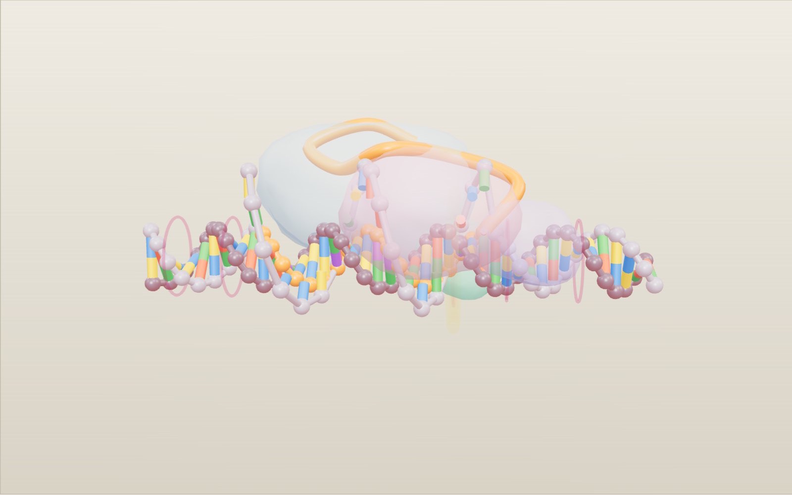
How CRISPR-Cas9 gene editing works
A guide RNA reads just 20 letters of DNA, and that is usually enough to pick out one spot among the 3 billion base pairs of a human genome.
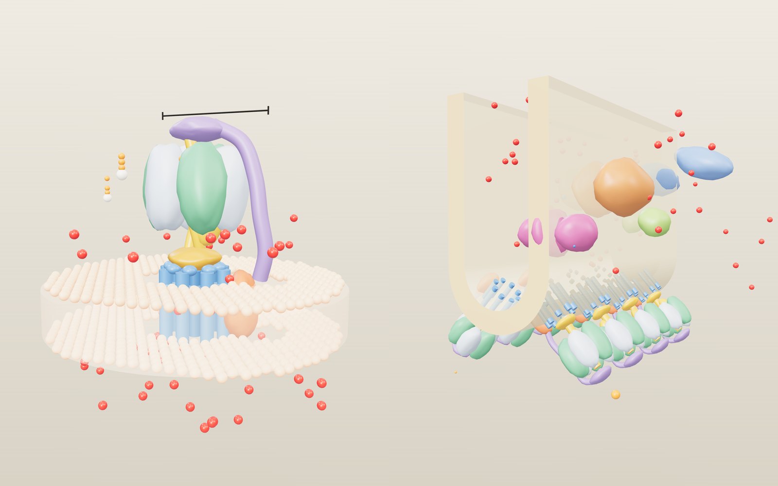
How ATP synthase works
In mitochondria the rotor of ATP synthase turns about 100 times a second, and every full turn releases three molecules of ATP.
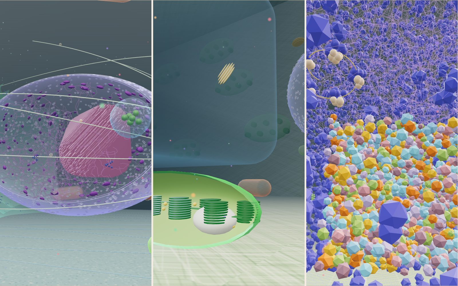
Walk inside a cell: animal, plant and bacterium
Grown a million times, a mitochondrion is about the size of a sofa, a whole E. coli is 2 m long, and the kinesin towing a vesicle past you walks at 0.8 metres per second.
How nerve signals travel
A nerve cell sits at about −70 mV, and when a stimulus lifts it to about −55 mV it fires a spike that is always the same size.
Giant sequoia forest: walk under the largest trees
Stand at the foot of the General Sherman Tree, 83.8 m tall and about 2,200 years old, then climb its trunk and see how hard the water up there is pulled.
Dinosaur size comparison to humans: 172 life-size animals
A 600 m park of 172 prehistoric animals at their real size, from the first dinosaurs to T. rex, laid out by age.