Loading the model
This device cannot show the 3D model. You can read the guide steps below as text.
1 / 11 84 base pairs of DNA, to scale This stretch is about 29 nm long. The bars below it give the scale: one turn is 10.5 base pairs (3.57 nm) in solution, while textbooks round it to 10 base pairs and 3.4 nm. The sequence is made up for the model.
Drag to turn. Scroll or pinch to zoom.
Guide Parts Controls
01 84 base pairs of DNA, to scale This stretch is about 29 nm long. The bars below it give the scale: one turn is 10.5 base pairs (3.57 nm) in solution, while textbooks round it to 10 base pairs and 3.4 nm. The sequence is made up for the model.
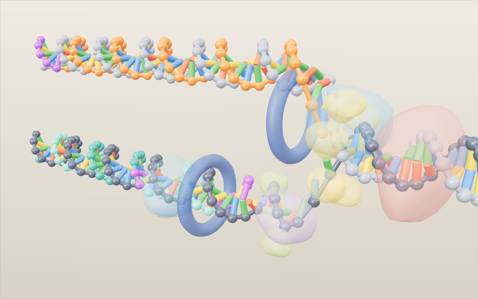
02 Helicase opens a fork The helicase ring pulls the strands apart and the DNA splits into a Y, the replication fork. Ahead of it a topoisomerase relieves the twist, and behind it small binding proteins (green) keep the single strands from closing again.
03 Every new piece starts with RNA DNA polymerase can only add to an existing 3′ end. Primase gets round this by writing a short RNA primer, the purple beads, on the lagging-strand template.
04 The leading strand grows in one piece On the top template the polymerase moves in the same direction as the fork, so it keeps adding to the same strand (orange) from one primer. Base colours are off here to show which strand is new.
05 The lagging strand comes in pieces The bottom new strand has to grow away from the fork. The polymerase builds one Okazaki fragment back to the previous one, lets go and starts again at the next primer, which leaves a row of pieces.
06 Primers out, nicks sealed DNA polymerase I replaces each RNA primer with DNA, starting from the fragment behind it. That leaves one missing bond, a nick, which DNA ligase seals. Watch the Okazaki count of joined fragments rise.
07 Taking back a wrong base Here the polymerase has put a G opposite a T. It stops, backs up one nucleotide, cuts the wrong base out and puts in the right one. Switch Proofreading off and the wrong base stays, while the fork gets ahead.
08 Pull the helicase yourself Drag the yellow helicase ring along the helix. The fork opens as fast as you pull, and Speed follows your hand. Pull backwards and nothing happens: a helicase moves only one way, using energy from ATP.
09 Faster fork, longer fragments Primase starts a new primer at regular moments, so the length of each Okazaki fragment depends on how far the fork has moved in between. In the model a primer comes every 2.8 seconds: 14 nt at 5 bp/s, 36 nt at 13 bp/s and 8 nt at 3 bp/s. In E. coli a new fragment starts every 1 to 2 seconds.
10 You are the polymerase Press Pair the next base. A white bead marks the next free place on the leading strand and a red ring the template base. Pick its partner: A for T, G for C. With Proofreading on a wrong pick is cut out again; with it off the mismatch stays.
11 Two copies, each half old When the fork has passed, there are two double helices. Each holds one old grey strand and one new coloured strand. Meselson and Stahl showed this in 1958 with heavy nitrogen, as the tubes in the corner sketch.
Parent DNA The two old strands, dark and light grey. Ahead of the fork they are still wound together, 2 nm across, one turn every 10.5 base pairs. Behind the fork each one ends up in a different copy. Base pairs A pairs with T and C with G, one pair every 0.34 nm. Colours: A green, T red, G yellow, C blue, U (in RNA) purple. Switch off Base pair colours to colour them by strand instead. Topoisomerase Works on the double helix ahead of the fork. Opening the strands overwinds the DNA in front; topoisomerase relieves the strain by cutting a strand and resealing it. Helicase Breaks the hydrogen bonds between the bases and pulls the strands apart, using energy from ATP. Drawn as a ring at the point where the strands part. Single-strand binding proteins Small proteins (green) that coat bare single strands near the fork so they do not pair up again before they are copied. They gather on the lagging-strand template, which waits longest. Primase An RNA polymerase that writes a short RNA primer on the template. DNA polymerase cannot start a strand on its own; it needs the primer's free 3′ end. RNA primers Short pieces of RNA (purple beads). One starts the leading strand; every Okazaki fragment on the lagging strand needs its own. Real primers are about 5 to 10 nucleotides; here they are 5. DNA polymerase and clamp Two polymerases (blue), one on each new strand, each held on the DNA by a ring-shaped sliding clamp. They add nucleotides only to a 3′ end, so each new strand grows 5′ to 3′. Leading strand The new strand (orange) whose 5′ to 3′ direction points towards the fork. It is made in one piece from a single primer and keeps pace with the helicase. Lagging strand, Okazaki fragments The other new strand (teal, two shades). Its 5′ to 3′ direction points away from the fork, so it is made backwards in pieces. Model fragments are 14 nucleotides; real ones are about 1,200 in bacteria and 200 in eukaryotes. DNA polymerase I In bacteria this enzyme removes each RNA primer and fills the gap with DNA, using the DNA of the next fragment as its starting point. In eukaryotes RNase H and other enzymes do this job. DNA ligase Seals the nick left between two pieces of DNA by forming the last phosphodiester bond. A flash marks each seal, and the fragments become one strand. Mismatch A wrong base the polymerase has put in. With Proofreading on, the polymerase stops, cuts it out with its 3′ to 5′ exonuclease and puts in the right one. With it off, the mismatch stays (ring).