FIG. 66How It Works
How nerve signals travel
A nerve cell sits at about −70 mV, and when a stimulus lifts it to about −55 mV it fires a spike that is always the same size.
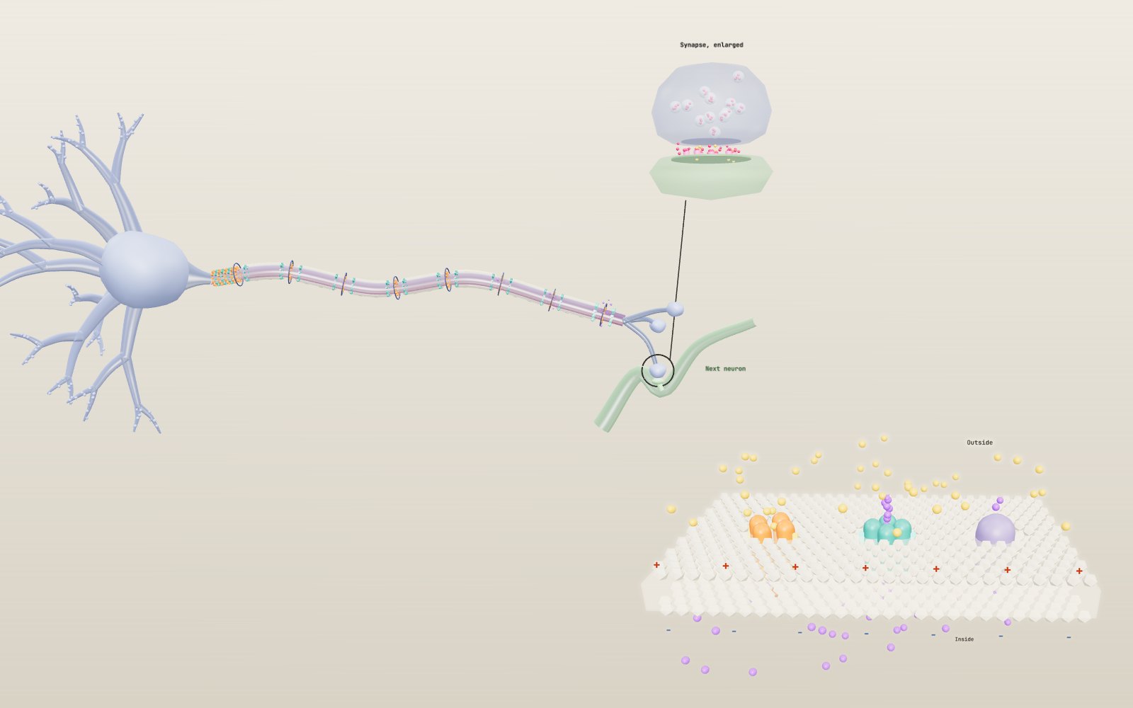
This device cannot show the 3D model. You can read the guide steps below as text.
1 / 28 One neuron from end to endSignals come in through the dendrites on the left and leave through the axon terminals on the right. The axon here is shortened and drawn far thicker than life. The panel in the corner plots the membrane potential at the axon hillock. The bar under the model opens seven more scenes.
Drag to turn. Scroll or pinch to zoom.
What happens when you stub your toe?
Stub a toe and you feel the knock first and the pain a moment later. Both messages travel up the same leg, along nerve fibres, to the spinal cord and brain. They arrive at different times because the fibres that carry them are built differently. Eric Chudler's Neuroscience for Kids pages at the University of Washington put the slow pain fibres at about 2.2 miles per hour, slower than you walk, while the thick touch fibres come close to the speed of an aeroplane.
Underneath that difference sits one mechanism that every nerve cell uses. A neuron keeps its inside electrically negative, and when a strong enough input arrives it flips that charge for about two milliseconds and passes the flip along its axon. At the far end the electrical signal becomes a chemical one that crosses to the next cell.
The exhibit opens on a single neuron in 3D. You can change the strength of the input, take the myelin off the axon, change its thickness and watch one synapse up close. The bar under the model switches to seven more scenes. One zooms in on the ion channels and holds the membrane at a fixed voltage, the way the experiments of the 1940s did. One races a myelinated fibre against a bare one over a metre. Others show five neurotransmitters at work, time a knee jerk from tap to kick, add up excitatory and inhibitory inputs and line up eight kinds of neuron side by side. The last is a squid giant axon in a dish, where the equations Alan Hodgkin and Andrew Huxley published in 1952 run live as you change the current, the temperature and the dose of two toxins. Under the controls are eight tasks to try and 51 short cases from the history of the subject.
A battery made of salt water
A neuron is a bag of salty water in a bath of different salty water. Inside there is a lot of potassium and little sodium; outside it is the other way round. OpenStax Biology gives typical values of 145 mM sodium outside and 12 mM inside, and 4 mM potassium outside against 155 mM inside. The sodium-potassium pump keeps it that way, pushing three sodium ions out and bringing two potassium ions in for every molecule of ATP it spends.
The membrane itself does not let ions through. They cross only through channel proteins, and at rest the open ones are mostly potassium channels. Potassium leaks out down its concentration gradient, each ion taking a positive charge with it, until the negative charge it leaves behind holds the rest back. The result, measured with one electrode inside the cell and one outside, is the resting membrane potential: about −70 mV. OpenStax notes that the exact figure varies between kinds of neuron and between species, but −70 mV is the number used in most textbooks and in this model.
In the exhibit, a blue membrane is a membrane at rest. The graph in the corner shows the potential at the axon hillock, and with the Stimulus slider at zero it stays flat on the −70 line.
Inside the channels
Ions cross the membrane through proteins, and the scene called Ion channels zooms in on three of them sitting in a square of lipid bilayer. The white beads are the head groups of the lipids, two sheets with an oily layer between them that no ion can cross on its own.
The orange protein is a voltage-gated sodium channel. It is one long chain folded into four similar domains round a central pore, a layout Shosaku Numa's group in Kyoto worked out in 1984 by reading the gene for the sodium channel of the electric eel. Each domain carries a voltage sensor, drawn as a yellow peg, that moves outward when the inside of the cell becomes less negative. On the inside hangs a loop drawn as a red ball on a chain. About a millisecond after the channel opens, that loop swings into the pore and plugs it. The channel cannot open again until the membrane has returned close to rest and the loop has swung back out.
The teal protein is a voltage-gated potassium channel, four separate subunits round a pore. Its sensors move more slowly, so it opens later than the sodium channel, and it has no fast plug. In 1998 Roderick MacKinnon's group solved the structure of a bacterial potassium channel and showed how a narrow filter lined with oxygen atoms lets potassium through while turning away sodium, which is smaller. That work shared the 2003 Nobel Prize in Chemistry.
The purple protein on the right is the sodium-potassium pump. Jens Skou found it in 1957 as an enzyme in crab nerve that split ATP only when both sodium and potassium were present, and he shared the 1997 Nobel Prize in Chemistry for it. For each ATP it moves three sodium ions out and two potassium ions in. The pump is slow next to the channels, and it does not make the action potential. Its job is to keep the concentration differences in place over minutes and hours.
Holding the voltage still
The scene runs a voltage clamp, the method Hodgkin and Huxley used. An amplifier measures the membrane potential and injects whatever current it takes to keep it at a chosen value. The current it has to supply is equal and opposite to the current flowing through the channels, so the clamp turns a fast, self-feeding event into something that can be measured one piece at a time.
The loop holds the membrane at −65 mV for 3 ms, steps it to the value of Clamp voltage for 12 ms, then returns. With the default step to −10 mV the graph shows two currents. Sodium current (orange, drawn downward because positive charge flows in) peaks at about 1,430 µA/cm² after 0.7 ms and then dies away, even though the voltage stays where it is. Potassium current (teal, upward) rises more slowly and is still flowing at about 1,430 µA/cm² when the step ends. The percentages in the readout give the open fraction of each kind of channel in the model, m³h for sodium and n⁴ for potassium.
Step to +50 mV and the sodium current vanishes, because +50 mV is the sodium equilibrium potential in the squid model: the channels open, but no net sodium moves. Step to +60 mV and the current reverses and flows outward. Hodgkin and Huxley used exactly this kind of reversal to prove which ion carried which current. The TTX and TEA sliders block a fraction of each kind of channel and change the two traces separately.
Inputs add up at the axon hillock
The dendrites are the receiving end. When transmitter from another neuron opens channels on a dendrite, a little positive charge flows in and the membrane there becomes less negative. These graded potentials are small and they fade as they spread, but many of them can arrive together and add up. OpenStax calls this summation, and it happens at the axon hillock, the cone where the axon leaves the cell body, together with the first stretch of the axon called the initial segment. That stretch is packed with voltage-gated sodium channels, which is why it is called the trigger zone.
Set the Stimulus slider to +10 mV and watch the yellow beads run down the dendrites. Each burst of input lifts the hillock to −60 mV and then sinks away again. Nothing travels down the axon.
Adding up in space and in time
A real neuron does not get one input at a time. A pyramidal cell in the cortex carries thousands of synapses on its dendrites, and some excite while others inhibit. The Summation scene has six excitatory synapses (green) on the dendrites and one inhibitory synapse (red) on the cell body next to the trigger zone, the first stretch of axon where the spike starts.
In the model each excitatory input adds 4 mV at its peak, rises in about 1 ms and fades with a time constant of 15 ms. Threshold sits 15 mV above the resting −70 mV, the same as in the neuron scene. Three inputs together reach −58 mV and fail. Four together reach −54 mV and the trigger zone fires. That is spatial summation: inputs at different places adding up because they arrive together.
Now spread them out. Input spacing sets the delay between one input and the next. With four inputs 2 ms apart, the first has already started to fade when the last arrives, and the peak stays at −55.7 mV, just short. Eight inputs can be spread up to 4.5 ms apart and still fire the cell. That is temporal summation, and it is why the timing of inputs matters as much as their number.
Inhibition changes the sum. Five simultaneous inputs would reach −50 mV, but an inhibitory input of 6 mV or more at the same moment keeps the cell below threshold. In the brain most fast inhibition comes from GABA opening chloride channels, and inhibitory synapses tend to sit on or near the cell body, where they cancel input from the whole dendritic tree. John Eccles recorded these inhibitory potentials in spinal motor neurons and shared the 1963 Nobel Prize with Hodgkin and Huxley.
The 4 mV per input is a model value chosen so that a handful of inputs is enough. A single synapse on a cortical neuron usually moves the cell body by well under 1 mV, so a real cell needs tens of inputs arriving close together.
Threshold and the all-or-none rule
Voltage-gated sodium channels open when the membrane reaches about −55 mV. That is the threshold. Once a few open, sodium rushes in, the membrane becomes less negative still, and more channels open. The loop runs away and the potential shoots up past zero to about +30 mV.
Two things then bring it back. Each sodium channel has a second gate that closes on its own within a fraction of a millisecond, so the inflow stops. At the same time voltage-gated potassium channels, which respond more slowly, open and let potassium out. The membrane falls back, overshoots to below −70 mV for a moment because the potassium channels are slow to close, and then settles. OpenStax puts the whole event at about 2 ms.
Below threshold none of this happens; at or above it all of it does, to the same height every time. Move the slider between +15 and +30 mV and the spike on the graph starts sooner but its top never moves. This is the all-or-none rule. A stronger stimulus does not make a bigger action potential. In real neurons it makes them come more often, and the model fires a little more often too as you push the slider up.
The squid axon and the 1952 equations
In 1936 the zoologist J. Z. Young showed that tubes running through the mantle of a squid, which had been taken for blood vessels, were single nerve fibres up to about a millimetre across. They drive the squid's jet escape, and they are wide enough to slide a wire inside. In 1939 Hodgkin and Huxley did just that at the Plymouth laboratory and recorded the potential from inside an axon for the first time. The spike did not stop at zero, as the membrane theory of Julius Bernstein predicted. It overshot zero by tens of millivolts.
After the war they used a voltage clamp with a silver wire threaded along the axon, so that the whole length behaved as one patch of membrane. They measured how sodium and potassium currents switched on and off at each voltage, and in 1952 they described them with a set of equations. Three gates, called m, h and n, open and close at rates that depend on voltage. Sodium conductance is 120 mS/cm² times m³h, potassium 36 mS/cm² times n⁴, and a small leak of 0.3 mS/cm² completes the circuit. The rates were measured at 6.3 °C, and the paper scales them by a factor of three for every 10 °C.
The Squid axon lab scene runs those equations as you watch. The grey trace in the readout is the potential recorded by the wire over 40 ms. The axon in the dish changes colour as the action potential travels along it, at the speed the same equations predict.
What the equations do
At 6.3 °C the model rests at −65 mV with the gates at m = 0.053, h = 0.596 and n = 0.318. A 1 ms pulse needs 6.9 µA/cm² to fire it. Below that the potential bumps up and sinks back. At 7 µA/cm² the spike comes late, about 5 ms after the pulse starts, and peaks near +35 mV. At 20 µA/cm² it comes after 1.3 ms and peaks near +41 mV, and doubling the current again adds less than 1 mV. Far above threshold the height hardly changes. Right at threshold the equations give a slightly smaller spike, a detail the textbook all-or-none rule smooths over. After the spike the membrane dips to about −76 mV, closer to the potassium equilibrium potential of −77 mV, because the potassium gates are still open.
Second pulse after sets a second pulse. With two pulses of 20 µA/cm² the second one fails if it comes less than 10.6 ms after the first. Double the current to 40 µA/cm² and a gap of 7.8 ms is enough. That is the relative refractory period: during recovery a stronger stimulus can fire the membrane earlier. Inside the first few milliseconds no pulse is strong enough, which is the absolute refractory period.
Steady current replaces the pulse with a step lasting 150 ms. From about 6.3 µA/cm² the axon fires again and again. At 10 µA/cm² it fires at 69 Hz, at 20 µA/cm² at 87 Hz and at 50 µA/cm² at 118 Hz. This is how a nerve cell turns strength into frequency, the code Edgar Adrian heard in sensory fibres in 1926.
Temperature speeds up all three gates together. At 18.5 °C they move 3.8 times faster, the spike is narrower and lower, peaking near +33 mV, and the threshold for a 1 ms pulse rises to 8.9 µA/cm². At 25 °C the peak is only about +19 mV, and from about 28 °C a 20 µA/cm² pulse no longer fires at all. Sodium inactivation and potassium activation then catch up with sodium activation before the spike can build.
How fast it travels
Part five of the 1952 paper did something more ambitious. Using the measured fibre radius of 238 µm and the resistance of the axoplasm, 35.4 Ω cm, Hodgkin and Huxley solved for an action potential that travels along the axon and found a speed of 18.8 m/s at 18.5 °C. The same axon had been measured at 21.2 m/s. Huxley did the arithmetic on a hand-cranked mechanical calculator.
The same calculation was repeated in a different way, by dividing a 6 cm axon into short pieces and stepping the whole cable forward in time. With pieces 0.5 mm long it gives 18.7 m/s, and with pieces of 0.25 mm the answer settles at 18.73 m/s, within half a per cent of the paper. At 6.3 °C the same cable conducts at 12.3 m/s. The lab uses that computed speed to colour the axon.
Two toxins
Tetrodotoxin (TTX), the poison of pufferfish, sticks in the outer mouth of voltage-gated sodium channels and plugs them. Toshio Narahashi and colleagues showed in 1964 on lobster giant axons that it removes the sodium current and leaves the potassium current alone. In the model a 20 µA/cm² pulse still fires with 71% of the sodium channels blocked and fails beyond that. At 100% the pulse pushes the membrane up and lets it fall back, and the wave along the axon disappears.
Tetraethylammonium (TEA) blocks potassium channels. In the model, block half of them and one pulse sets off a short burst of three spikes because nothing pulls the membrane down firmly. Block 90% and the resting potential drifts up until the sodium channels sit inactivated and nothing fires. Both toxins are shown here as tools that physiologists use. They are dangerous substances, and the exhibit says nothing about them as medicines or treatments.
Why the signal moves only one way
A spike at the hillock pushes positive charge along the inside of the axon. That charge lifts the next patch of membrane to threshold, its sodium channels open, and the patch fires in turn. The patch behind cannot fire again straight away because its sodium channels are still inactivated. This is the absolute refractory period, and it is why the action potential runs toward the terminals and never back toward the cell body.
On a bare axon every patch has to do this in turn. The result is a continuous wave, and it is slow. Switch off Myelin in Controls and watch the amber band crawl along the axon.
Myelin and the nodes of Ranvier
Most fast axons in vertebrates are wrapped in myelin, layer upon layer of glial cell membrane. In nerves outside the brain and spinal cord each wrap comes from a Schwann cell; inside the brain and spinal cord oligodendrocytes make it, one cell sending out arms to several axons. The sheath insulates the axon, so current spreads along the inside instead of leaking out through the membrane.
The sheath is not continuous. Between the wraps are gaps about one micrometre long, the nodes of Ranvier, and that is where the voltage-gated sodium channels sit. A spike at one node drives current down the insulated stretch fast enough to bring the next node to threshold, and the action potential is rebuilt there. It seems to jump from node to node, which gave it the name saltatory conduction, from the Latin for leaping. In the model the nodes light up one after another while the stretches between them glow only faintly.
The gain is large. In Purves and colleagues' Neuroscience, unmyelinated axons conduct at about 0.5 to 10 m/s while myelinated axons can reach 150 m/s. Thickness matters too. A wider axon has less internal resistance, so the spread of charge reaches farther. Change Axon diameter and the speed in the corner changes with it, faster for thick fibres and slower for thin ones.
Two ways to go fast
The Myelin race scene puts two axons side by side over one metre. Both start at 10 µm. The myelinated one finishes in 17.5 ms and the bare one in 316 ms, 18 times longer. The glow on the upper fibre hops from node to node; only twelve sheaths are drawn, although a 10 µm fibre would have about a thousand in a metre, each about 1 mm long and crossed in about 18 µs.
Animals have found two answers to the problem of speed. Vertebrates wrap their fast axons in myelin. Squid, and many other invertebrates, make the axon wider instead, because a wider core has less resistance and the spread of current reaches further. Set Bare axon width to 476 µm, the size of Hodgkin and Huxley's axon, and the bare fibre reaches 21.8 m/s on the model's square-root rule, close to the 21.2 m/s they measured. It is 50 times wider than the myelinated fibre beside it and still nearly three times slower. A myelinated fibre needs only about 4 µm to reach 21 m/s, a cross-section more than ten thousand times smaller, which is one reason a human nerve can carry thousands of fast fibres.
The fastest fibres on record are invertebrate ones with myelin of their own. Ke Xu and Susumu Terakawa reviewed the giant fibres of Penaeus shrimp, which conduct at more than 200 m/s. Their myelin leaves a wide space between itself and the axon, and instead of ordinary nodes it has small windows.
Damaged myelin removes the sheath from a stretch in the middle of the fibre. The model treats that stretch as a bare axon, so the signal slows there and finishes later. In real nerves the current can leak away through the exposed membrane, which has few sodium channels, and the signal can fail altogether. Diseases in which the immune system attacks myelin, such as multiple sclerosis in the brain and spinal cord or Guillain-Barré syndrome in peripheral nerves, slow or block conduction in this way. The scene illustrates the idea only. NINDS, part of the US National Institutes of Health, keeps patient information on both.
Checking the numbers
The resting potential is close to the equilibrium potential of potassium, and the Nernst equation gives that value from the two concentrations:
E = (RT / zF) × ln(outside / inside)
At body temperature, 37 °C, RT/F is 26.73 mV, which comes to 61.54 mV for each tenfold difference in concentration. For potassium, 61.54 × log₁₀(4/155) gives −97.7 mV. For sodium, 61.54 × log₁₀(145/12) gives +66.6 mV. For chloride, with its negative charge, the same numbers give −90.9 mV.
The resting value of −70 mV lies between them because the membrane at rest lets a little sodium in as well. The Goldman equation weighs each ion by how easily it crosses. If sodium crosses at one twentieth of the rate of potassium (chloride left out), the formula gives −70.2 mV. Make the ratio one hundredth and it falls to −89.5 mV; raise it to one tenth and it climbs to −57.0 mV. The peak of +30 mV stays well short of sodium's +66.6 mV, because the sodium channels inactivate and the potassium channels open before it can get there.
For conduction speed the model uses two rules. A myelinated fibre conducts at about 5.7 m/s for each micrometre of diameter, a rule of thumb from the Washington University Neuromuscular Disease Center. A bare axon is given 1 m/s at 1 µm, matching thin pain fibres, rising with the square root of diameter. At 10 µm that makes 57 m/s with myelin and 3.16 m/s without, a factor of 18. Over one metre of axon the signal takes 17.5 ms in the first case and 316 ms in the second. At 20 µm the myelinated fibre reaches 114 m/s, and across the whole slider the values stay inside the textbook ranges.
The same source gives an internode length of about 100 times the axon diameter, so about 1 mm for a 10 µm fibre. At 57 m/s that is 17.5 µs from node to node, the same order as the 25 µs it quotes. The threshold in the model is reached once the input adds 15 mV: +20 mV crosses it after 0.42 ms, +30 mV after 0.23 ms. All of these figures were also worked through numerically.
From electricity to chemistry at the synapse
At the end of the axon the signal has to cross a gap to the next cell. When the action potential reaches a bouton, the swelling at the tip of each terminal branch, voltage-gated calcium channels open and calcium flows in. Calcium makes the synaptic vesicles that are docked at the membrane fuse with it and spill their neurotransmitter into the synaptic cleft. The transmitter diffuses across and binds receptor proteins on the next neuron. Many of those receptors are themselves ion channels, so binding opens them and positive ions flow in, nudging that cell toward its own threshold. Then the transmitter is broken down, drifts away or is pumped back in, and the synapse is ready again.
OpenStax gives about one millisecond between the action potential arriving and the channels opening on the other side. The close-up in the model shows calcium in green, transmitter in pink and receptors that glow while transmitter is bound to them.
Five transmitters
The Synapse scene enlarges one synapse until the vesicles, the cleft and the receptors fill the stage. Transmitter switches between five of the best known neurotransmitters, and each one changes the receptors, the response and the way the cleft is cleared.
- Glutamate is the main fast excitatory transmitter of the brain. Most of its receptors are ion channels (AMPA and NMDA receptors) that let positive ions in within a millisecond, giving a fast excitatory postsynaptic potential (EPSP). Transporters, many of them on astrocytes, pull it out of the cleft.
- GABA is the main inhibitory transmitter. GABA-A receptors are chloride channels, and the response is an inhibitory postsynaptic potential (IPSP) that holds the cell away from threshold. GABA is made from glutamate and cleared by its own transporters.
- Acetylcholine carries the signal from motor neurons to skeletal muscle through nicotinic receptors, which are ion channels. It is not taken back up whole. The enzyme acetylcholinesterase in the cleft splits it, and the choline is recycled. Otto Loewi's experiment with two frog hearts in 1921 first showed a nerve releasing a chemical, which turned out to be acetylcholine.
- Dopamine acts only through receptors coupled to G proteins (D1 to D5). The response starts later, is smaller and lasts longer, and its main effect is to change how the target cell answers its other inputs. Neurons in the midbrain send it to the striatum and the cortex, and the dopamine transporter clears it.
- Serotonin is made from tryptophan by neurons of the raphe nuclei, which reach most of the brain. Almost all its receptors are G-protein-coupled; the 5-HT3 receptor is the exception, an ion channel. The serotonin transporter clears it.
Reuptake switches the transporters off. The molecules then stay in the cleft longer and the response stretches out. Some medicines act on this step by slowing a transporter. The scene shows the principle only.
The cleft is about 20 nm across, so diffusion across it takes microseconds. Most of the roughly 1 ms synaptic delay goes into calcium entry and vesicle fusion. Bernard Katz and José del Castillo showed in 1954 at the frog nerve-muscle junction that transmitter comes out in packets of fixed size, later matched to single vesicles. Katz shared the 1970 Nobel Prize with Ulf von Euler, who identified noradrenaline as a transmitter, and Julius Axelrod, who showed that it is taken back into the nerve ending after release.
The knee jerk, timed
A doctor's tap below the kneecap stretches the thigh muscle for an instant. Stretch receptors inside the muscle, the muscle spindles, fire, and the signal runs up thick sensory fibres (called Ia fibres) through the dorsal root into the spinal cord. There each fibre makes a synapse directly on the motor neurons of the same muscle, and their axons carry the command straight back down. The muscle contracts and the leg kicks. Only one synapse lies on the path, which is why the reflex is so quick. At the same time a branch of the sensory fibre excites an inhibitory interneuron that relaxes the hamstrings on the back of the thigh. Charles Sherrington worked out this arrangement, reciprocal inhibition, and coined the word synapse; he shared the 1932 Nobel Prize with Adrian.
The Knee jerk scene times the trip. The model takes the one-way path from muscle to spinal cord as a share of height: 0.29 of height for the knee, whose motor neurons sit in the L3 and L4 segments of the cord, and 0.52 for the calf, served by S1. It sends the signal in at 65 m/s and out at 55 m/s, values inside the range of large myelinated fibres, and adds three fixed delays of 1 ms for the spindle, the central synapse and the nerve-muscle junction. For a person 175 cm tall that is 0.51 m each way, 7.8 ms in and 9.2 ms out, 20.0 ms in all.
Measurements agree. Helen Vickery and Paul Smith at the University of Nottingham recorded the patellar reflex with surface electrodes on the thigh of 28 young adults and found 17 ms in women and 21 ms in men. C. J. Frijns and colleagues in Amsterdam measured knee and ankle reflexes in 102 healthy people and found that latency grows with height, with a correlation of 0.70 for the knee and 0.72 for the ankle. Each person's latency hardly changed from tap to tap.
Set Tap at to Ankle. The path from the calf is almost twice as long, and the model gives 33.5 ms at 175 cm. The Washington University Neuromuscular Disease Center gives about 30 ms for the soleus H reflex, an electrical version of the ankle jerk that starts the signal in the nerve instead of the spindle. Change Height from 140 cm to 200 cm and the knee jerk goes from 16.6 ms to 22.5 ms.
None of this involves the brain, which is why a knee jerk is faster than any reaction you can make on purpose. A reaction you choose to make takes a few hundred milliseconds, and the reaction time exhibit measures yours.
Many shapes of neuron
Santiago Ramón y Cajal spent decades drawing neurons stained with Camillo Golgi's silver method and showed that they are separate cells with recognisable shapes. The Neuron types scene lines up eight of them, all drawn at the same height so the branching can be compared. The readout gives each one's real size.
- Pyramidal cells are the main excitatory neurons of the cerebral cortex. A long apical dendrite climbs toward the surface and ends in a tuft, while basal dendrites spread round the base. They release glutamate.
- Purkinje cells of the cerebellum spread their dendrites in a flat fan. Parallel fibres cross the fan at right angles; Richard Napper and Robert Harvey counted about 175,000 parallel fibre synapses on one Purkinje cell in the rat. The cells are inhibitory and send their axons to the deep cerebellar nuclei.
- Motor neurons of the spinal cord have large cell bodies, long dendrites and a thick myelinated axon that can reach a muscle a metre away.
- Retinal bipolar cells have one process up to the rods or cones and one down to the ganglion cells. Most types pass graded potentials instead of spikes, and the retina has more than ten types.
- Cerebellar granule cells are among the smallest neurons, with four short claw-like dendrites and an axon that climbs and splits into two parallel fibres. Frederico Azevedo and colleagues counted about 86 billion neurons in the human brain, and about four in five of them are in the cerebellum, mostly granule cells.
- Sensory neurons of the dorsal root ganglia are pseudo-unipolar: one stem leaves the cell body and splits, one branch running to the skin or muscle and the other into the spinal cord. The signal can pass the junction without going through the cell body.
- Chandelier cells are inhibitory interneurons of the cortex. Their boutons stand in short vertical rows like candles, and Péter Somogyi showed in 1977 that each row ends on the initial segment of a pyramidal cell's axon, the place where spikes begin.
- Medium spiny neurons make up most of the striatum. They are inhibitory, densely covered in spines and receive both cortical glutamate and midbrain dopamine.
The shapes in the scene are simplified drawings. NeuroMorpho.Org, an archive started by Giorgio Ascoli's group at George Mason University, collects reconstructions of real neurons traced from microscope images by many laboratories, and is the place to compare their shapes in detail.
What the model leaves out
Nothing in the drawing is to scale. The axon is far shorter and fatter than any real one, and the synapse is enlarged into a separate close-up. Time is slowed so that one millisecond lasts half a second on screen. The spike on the screen does not move at the true relative speeds either. A myelinated axon is 18 times faster than the same axon bare, and the model narrows that gap so you do not wait half a minute, but the readout always gives the real figures.
In the neuron scene the shape of the action potential is a drawn curve through the textbook values of −70, −55 and +30 mV, not a simulation of channel kinetics. The squid lab is the place where real kinetics run, and its numbers belong to the squid axon at its own temperature, with rest at −65 mV and a peak near +40 mV. Mammalian neurons have more kinds of channel, and their spikes are shorter.
The other scenes simplify as well. The ion channels are shaped after their known structure but drawn as smooth blocks, and the pump turns far more slowly than a real one. The race uses two rules of thumb for speed and draws twelve internodes. Synaptic responses in the synapse scene are model shapes. The reflex model takes path length as a fixed share of height and leaves out the time a muscle needs to start moving after its electrical signal. In the summation scene each input is far stronger than a real one. The neuron types are drawn at one size.
This exhibit explains how nerve cells work in general. It is not medical advice.
Who worked it out
Luigi Galvani made frog legs twitch with metal arcs in the 1780s and argued for animal electricity. In 1850 Hermann von Helmholtz timed a frog nerve at about 27 m/s, slow enough to show that nerve signals are not electricity in a wire. Julius Bernstein proposed in 1902 that the resting membrane is permeable to potassium.
Cajal and Golgi shared the 1906 Nobel Prize for the structure of the nervous system. Sherrington and Adrian shared the 1932 prize, Sherrington for reflexes and synapses, Adrian for the all-or-none spike and frequency coding. Henry Dale and Otto Loewi shared the 1936 prize for chemical transmission. Joseph Erlanger and Herbert Gasser won in 1944 for showing that thicker fibres conduct faster. Ichiji Tasaki in 1939, and Andrew Huxley with Robert Stämpfli in 1949, showed that excitation in myelinated fibres jumps between the nodes.
Alan Hodgkin and Andrew Huxley measured the sodium and potassium currents in the giant axon of the squid and described them with equations in 1952. They shared the 1963 Nobel Prize in Physiology or Medicine with John Eccles, who studied synapses in the central nervous system. The prize was given for the ionic mechanisms of excitation and inhibition in the nerve cell membrane.
Later work found the molecules behind the equations. Erwin Neher and Bert Sakmann sealed a fine glass pipette onto a muscle cell in 1976 and recorded the current through single channels, and shared the 1991 prize. The genes and then the structures of the channels followed in the 1980s and 1990s. Arvid Carlsson, Paul Greengard and Eric Kandel shared the 2000 prize for signalling between nerve cells, including Carlsson's work on dopamine.
Questions
What is an action potential?
A brief reversal of the voltage across a neuron's membrane, from about −70 mV to about +30 mV and back, lasting about 2 ms. It starts at the axon hillock and travels along the axon to the terminals.
Why is the inside of a neuron negative at rest?
At rest the membrane lets potassium through far more easily than sodium. Potassium leaks out down its concentration gradient and leaves the inside negative, and the sodium-potassium pump keeps the concentrations from running down.
If every spike is the same size, how does a neuron signal a strong stimulus?
By firing more often. A stronger input reaches threshold sooner and starts the next spike earlier, so the frequency of action potentials carries the strength while each one stays the same height.
How fast do nerve signals travel?
It depends on myelin and thickness. Unmyelinated axons conduct at about 0.5 to 10 m/s and myelinated ones at up to about 150 m/s, according to Purves and colleagues. The thinnest pain fibres manage about 1 m/s.
What does myelin do?
It insulates the axon so that the action potential only has to be rebuilt at the nodes of Ranvier. That makes conduction much faster and saves energy, since the channels and pumps are concentrated at the nodes. In demyelinating diseases such as multiple sclerosis, current leaks out and conduction slows.
Why does the signal not travel backwards?
Behind the spike, the sodium channels are inactivated for a short time and cannot open again. This refractory period stops the action potential from turning round, so it runs from the cell body toward the terminals.
How long does it take to cross a synapse?
About one millisecond at a chemical synapse, for calcium entry, vesicle fusion, diffusion across the cleft and opening of the receptors. Electrical synapses, where the cells are joined by channels, are almost instantaneous.
Is the resting potential always −70 mV?
No. It varies with the type of neuron and the species, and textbooks use −70 mV as a typical value. The threshold of about −55 mV and the peak of +30 to +40 mV are typical values too.
What did Hodgkin and Huxley actually discover?
That the action potential is carried by two separate currents: sodium flowing in first, then potassium flowing out. They measured how each switched on and off with voltage and time, and wrote equations that reproduced the shape of the spike, the threshold, the refractory period and the speed of conduction in the squid axon.
What is a voltage clamp?
An amplifier that holds the membrane potential at a chosen value by injecting whatever current is needed. The injected current equals the current through the channels, so it can be measured. The Ion channels scene runs a clamp loop.
Why does a squid have such a thick axon?
The giant axons drive the squid's escape jet, and a wider axon conducts faster because its core has less resistance. A 0.5 mm axon reaches about 21 m/s without myelin, which vertebrates get from a myelinated fibre about 4 µm across.
Why is the knee jerk so fast?
Only one synapse lies between the sensory fibre and the motor neuron, and both fibres are thick and myelinated. The brain is not involved. The whole trip takes about 17 to 21 ms in young adults, longer in taller people.
What is the difference between excitatory and inhibitory synapses?
An excitatory synapse pushes the next cell toward threshold, usually by letting positive ions in. An inhibitory synapse holds it away, usually by letting chloride in. The trigger zone fires only if the sum of all inputs reaches threshold.
What do toxins such as TTX do to nerves?
Tetrodotoxin blocks voltage-gated sodium channels, so action potentials cannot be generated or conducted. This exhibit shows the effect in a model to explain how channels work. It is not information about poisoning or treatment.
Terms
- Membrane potential: the voltage across the cell membrane, inside measured against outside.
- Resting potential: the steady membrane potential of a neuron that is not signalling, about −70 mV.
- Threshold: the potential, about −55 mV, at which voltage-gated sodium channels open and an action potential starts.
- Depolarization: a change that makes the inside less negative.
- Repolarization: the return toward the resting potential after a spike.
- Hyperpolarization: a dip below the resting potential, as after an action potential.
- Refractory period: the time after a spike when the membrane cannot fire again, or only with a stronger stimulus.
- Axon hillock and initial segment: the trigger zone where the axon leaves the cell body.
- Myelin: insulating wraps of glial membrane around an axon.
- Node of Ranvier: a short gap between myelin wraps where the action potential is rebuilt.
- Saltatory conduction: conduction that jumps from node to node along a myelinated axon.
- Synaptic vesicle: a small membrane sac filled with neurotransmitter.
- Nernst equation: the formula for the voltage at which one kind of ion is in balance across a membrane.
- Voltage clamp: a method that holds the membrane at a set voltage and measures the current through its channels.
- Gating variables (m, h, n): the open fractions of the sodium activation, sodium inactivation and potassium gates in the Hodgkin-Huxley model.
- Spatial summation: inputs at different places adding up because they arrive together.
- Temporal summation: inputs at one place adding up because they arrive close together in time.
- EPSP and IPSP: excitatory and inhibitory postsynaptic potentials, small changes that push a cell toward or away from threshold.
- Ionotropic receptor: a receptor that is itself an ion channel.
- Metabotropic receptor: a receptor that acts through a G protein inside the cell.
- Reuptake: removal of transmitter from the cleft by transporter proteins.
- Muscle spindle: a stretch receptor inside a muscle.
- Reflex arc: the path from receptor to spinal cord to muscle that produces a reflex.
- Reciprocal inhibition: relaxation of the opposing muscle during a reflex, through an inhibitory interneuron.
Sources
- OpenStax, Anatomy and Physiology 2e, 12.4 The Action Potential (−70 mV rest, −55 mV threshold, +30 mV peak, about 2 ms, sodium inactivation gate, refractory period, saltatory conduction). https://openstax.org/books/anatomy-and-physiology-2e/pages/12-4-the-action-potential (checked 16 October 2026)
- OpenStax, Biology 2e, 35.2 How Neurons Communicate (ion concentrations in Table 35.1, −70 mV varies by neuron and species, peak about +40 mV, nodes about 1 µm, pump 3 Na⁺ out and 2 K⁺ in, synaptic delay about 1 ms, multiple sclerosis). https://openstax.org/books/biology-2e/pages/35-2-how-neurons-communicate (checked 16 October 2026)
- OpenStax, Anatomy and Physiology 2e, 12.2 Nervous Tissue (axon hillock, initial segment and trigger zone, synaptic end bulbs, Schwann cells and oligodendrocytes). https://openstax.org/books/anatomy-and-physiology-2e/pages/12-2-nervous-tissue (checked 16 October 2026)
- OpenStax, Anatomy and Physiology 2e, 12.5 Communication Between Neurons (calcium and vesicle fusion, synaptic cleft, summation at the axon hillock). https://openstax.org/books/anatomy-and-physiology-2e/pages/12-5-communication-between-neurons (checked 16 October 2026)
- Purves D et al., Neuroscience, 2nd edition, Increased Conduction Velocity as a Result of Myelination, NCBI Bookshelf (unmyelinated 0.5 to 10 m/s, myelinated up to 150 m/s, saltatory conduction). https://www.ncbi.nlm.nih.gov/books/NBK10921/ (checked 16 October 2026)
- Chudler EH, Neuroscience for Kids, Conduction Velocity, University of Washington (fibre groups, C fibres about 2.2 miles per hour). https://faculty.washington.edu/chudler/cv.html (checked 16 October 2026)
- Washington University Neuromuscular Disease Center, Peripheral axons: characteristics (velocity about 5.7 × diameter, g-ratio 0.6 to 0.7, internode about 100 × diameter, about 25 µs between nodes). https://neuromuscular.wustl.edu/nother/axon.htm (checked 16 October 2026)
- Hodgkin AL, Huxley AF. A quantitative description of membrane current and its application to conduction and excitation in nerve. Journal of Physiology 117, 500 (1952). https://pmc.ncbi.nlm.nih.gov/articles/PMC1392413/ (checked 16 October 2026)
- NobelPrize.org, The Nobel Prize in Physiology or Medicine 1963, summary (Eccles, Hodgkin, Huxley). https://www.nobelprize.org/prizes/medicine/1963/summary/ (checked 16 October 2026)
- NobelPrize.org, The Nobel Prize in Physiology or Medicine 1936, summary (Dale, Loewi, chemical transmission). https://www.nobelprize.org/prizes/medicine/1936/summary/ (checked 16 October 2026)
- Hodgkin AL, Huxley AF. Action potentials recorded from inside a nerve fibre. Nature 144, 710 (1939). doi:10.1038/144710a0 (checked 18 October 2026)
- Young JZ. Structure of nerve fibres and synapses in some invertebrates. Cold Spring Harbor Symposia on Quantitative Biology 4, 1 (1936). doi:10.1101/SQB.1936.004.01.001 (checked 18 October 2026)
- Narahashi T, Moore JW, Scott WR. Tetrodotoxin blockage of sodium conductance increase in lobster giant axons. Journal of General Physiology 47, 965 (1964). doi:10.1085/jgp.47.5.965 (checked 18 October 2026)
- Noda M et al. Primary structure of Electrophorus electricus sodium channel deduced from cDNA sequence. Nature 312, 121 (1984). doi:10.1038/312121a0 (checked 18 October 2026)
- Doyle DA et al. The structure of the potassium channel: molecular basis of K⁺ conduction and selectivity. Science 280, 69 (1998). doi:10.1126/science.280.5360.69 (checked 18 October 2026)
- Skou JC. The influence of some cations on an adenosine triphosphatase from peripheral nerves. Biochimica et Biophysica Acta 23, 394 (1957). doi:10.1016/0006-3002(57)90343-8 (checked 18 October 2026)
- Neher E, Sakmann B. Single-channel currents recorded from membrane of denervated frog muscle fibres. Nature 260, 799 (1976). doi:10.1038/260799a0 (checked 18 October 2026)
- del Castillo J, Katz B. Quantal components of the end-plate potential. Journal of Physiology 124, 560 (1954). doi:10.1113/jphysiol.1954.sp005129 (checked 18 October 2026)
- Adrian ED, Zotterman Y. The impulses produced by sensory nerve-endings, part 2. Journal of Physiology 61, 151 (1926). doi:10.1113/jphysiol.1926.sp002281 (checked 18 October 2026)
- Tasaki I. The electro-saltatory transmission of the nerve impulse and the effect of narcosis upon the nerve fiber. American Journal of Physiology 127, 211 (1939). doi:10.1152/ajplegacy.1939.127.2.211 (checked 18 October 2026)
- Huxley AF, Stämpfli R. Evidence for saltatory conduction in peripheral myelinated nerve fibres. Journal of Physiology 108, 315 (1949). doi:10.1113/jphysiol.1949.sp004335 (checked 18 October 2026)
- Hursh JB. Conduction velocity and diameter of nerve fibers. American Journal of Physiology 127, 131 (1939). doi:10.1152/ajplegacy.1939.127.1.131 (checked 18 October 2026)
- Xu K, Terakawa S. Fenestration nodes and the wide submyelinic space form the basis for the unusually fast impulse conduction of shrimp myelinated axons. Journal of Experimental Biology 202, 1979 (1999). doi:10.1242/jeb.202.15.1979 (checked 18 October 2026)
- Vickery HSF, Smith PA. Surface electromyography reveals males have a slower patellar reflex than females. Journal of Electromyography and Kinesiology 22, 990 (2012). doi:10.1016/j.jelekin.2012.06.001 (checked 18 October 2026)
- Frijns CJM, Laman DM, van Duijn MAJ, van Duijn H. Normal values of patellar and ankle tendon reflex latencies. Clinical Neurology and Neurosurgery 99, 31 (1997). doi:10.1016/s0303-8467(96)00593-8 (checked 18 October 2026)
- Washington University Neuromuscular Disease Center, Tendon reflexes (H reflex latency about 30 ms, rises with height). https://neuromuscular.wustl.edu/mother/reflex.html (checked 18 October 2026)
- Napper RMA, Harvey RJ. Number of parallel fiber synapses on an individual Purkinje cell in the cerebellum of the rat. Journal of Comparative Neurology 274, 168 (1988). doi:10.1002/cne.902740204 (checked 18 October 2026)
- Azevedo FAC et al. Equal numbers of neuronal and nonneuronal cells make the human brain an isometrically scaled-up primate brain. Journal of Comparative Neurology 513, 532 (2009). doi:10.1002/cne.21974 (checked 18 October 2026)
- Spruston N. Pyramidal neurons: dendritic structure and synaptic integration. Nature Reviews Neuroscience 9, 206 (2008). doi:10.1038/nrn2286 (checked 18 October 2026)
- Euler T et al. Retinal bipolar cells: elementary building blocks of vision. Nature Reviews Neuroscience 15, 507 (2014). doi:10.1038/nrn3783 (checked 18 October 2026)
- Somogyi P. A specific axo-axonal interneuron in the visual cortex of the rat. Brain Research 136, 345 (1977). doi:10.1016/0006-8993(77)90808-3 (checked 18 October 2026)
- Gerfen CR, Surmeier DJ. Modulation of striatal projection systems by dopamine. Annual Review of Neuroscience 34, 441 (2011). doi:10.1146/annurev-neuro-061010-113641 (checked 18 October 2026)
- Ascoli GA, Donohue DE, Halavi M. NeuroMorpho.Org: a central resource for neuronal morphologies. Journal of Neuroscience 27, 9247 (2007). doi:10.1523/JNEUROSCI.2055-07.2007 (checked 18 October 2026)
- Korn H, Faber DS. The Mauthner cell half a century later. Neuron 47, 13 (2005). doi:10.1016/j.neuron.2005.05.019 (checked 18 October 2026)
- Piccolino M. Animal electricity and the birth of electrophysiology: the legacy of Luigi Galvani. Brain Research Bulletin 46, 381 (1998). doi:10.1016/S0361-9230(98)00026-4 (checked 18 October 2026)
- Schmidgen H. Of frogs and men: the origins of psychophysiological time experiments, 1850 to 1865. Endeavour 26, 142 (2002). doi:10.1016/S0160-9327(02)01466-7 (checked 18 October 2026)
- Seyfarth EA. Julius Bernstein (1839 to 1917): pioneer neurobiologist and biophysicist. Biological Cybernetics 94, 2 (2006). doi:10.1007/s00422-005-0031-y (checked 18 October 2026)
- NobelPrize.org, The Nobel Prize in Physiology or Medicine 1906, 1932, 1944, 1970, 1991 and 2000, and in Chemistry 1997 and 2003, summaries. https://www.nobelprize.org/prizes/medicine/1991/summary/ (checked 18 October 2026)
- National Institute of Neurological Disorders and Stroke (NINDS), Multiple sclerosis; Guillain-Barré syndrome; Myasthenia gravis; Parkinson's disease; Amyotrophic lateral sclerosis. https://www.ninds.nih.gov/health-information/disorders/multiple-sclerosis (checked 18 October 2026)
More exhibits
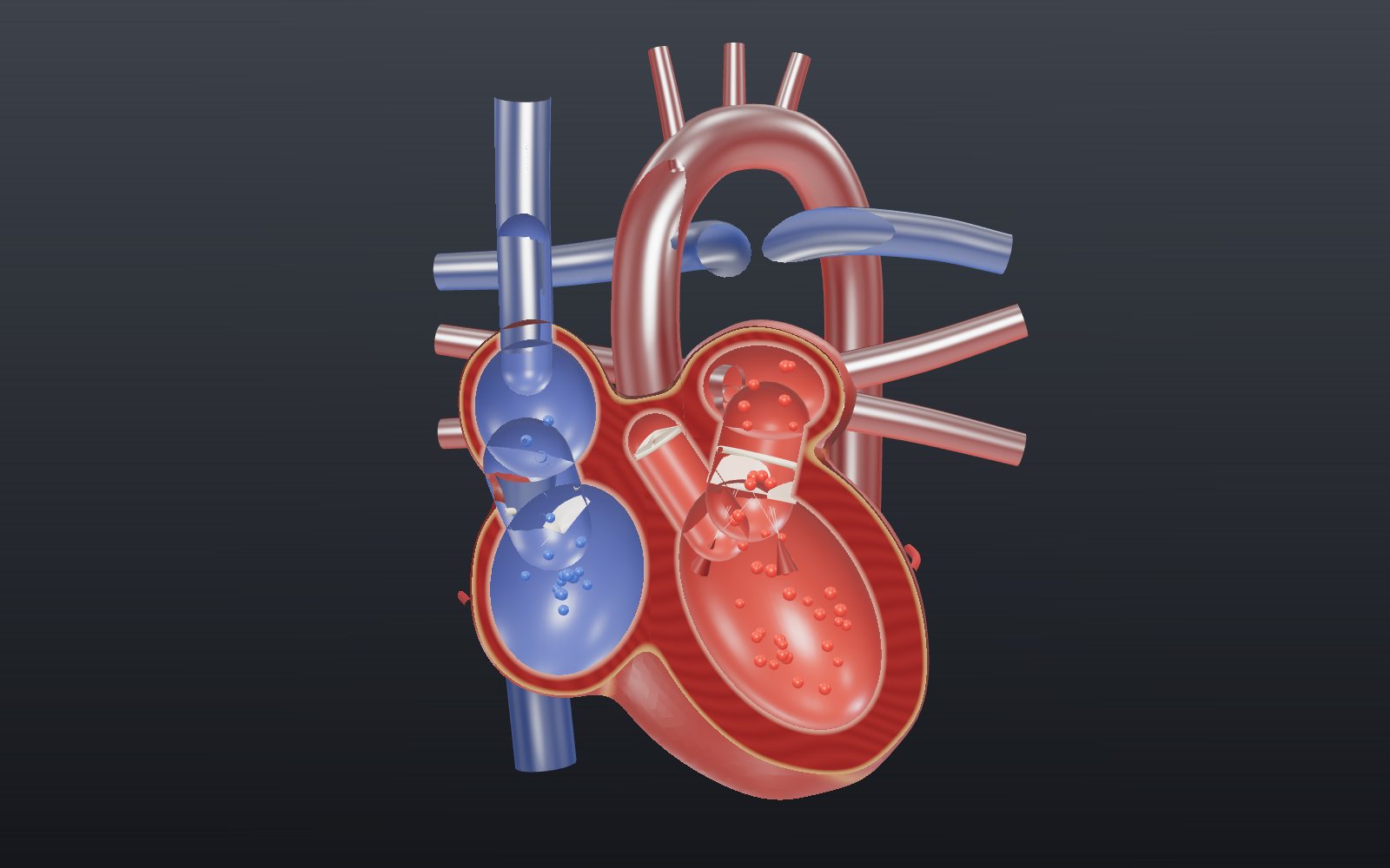
Heart beating: interactive model of the heart, step by step
Your heart pushes out about 70 mL of blood with each beat, and at a resting pace it does that about 108,000 times a day.
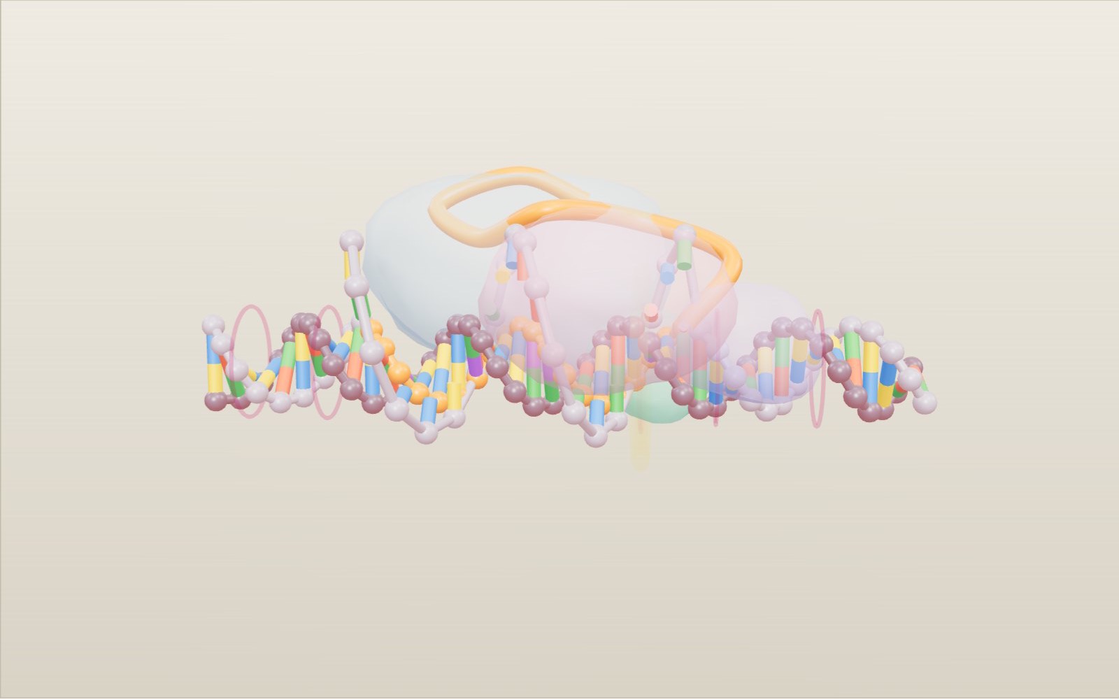
How CRISPR-Cas9 gene editing works
A guide RNA reads just 20 letters of DNA, and that is usually enough to pick out one spot among the 3 billion base pairs of a human genome.
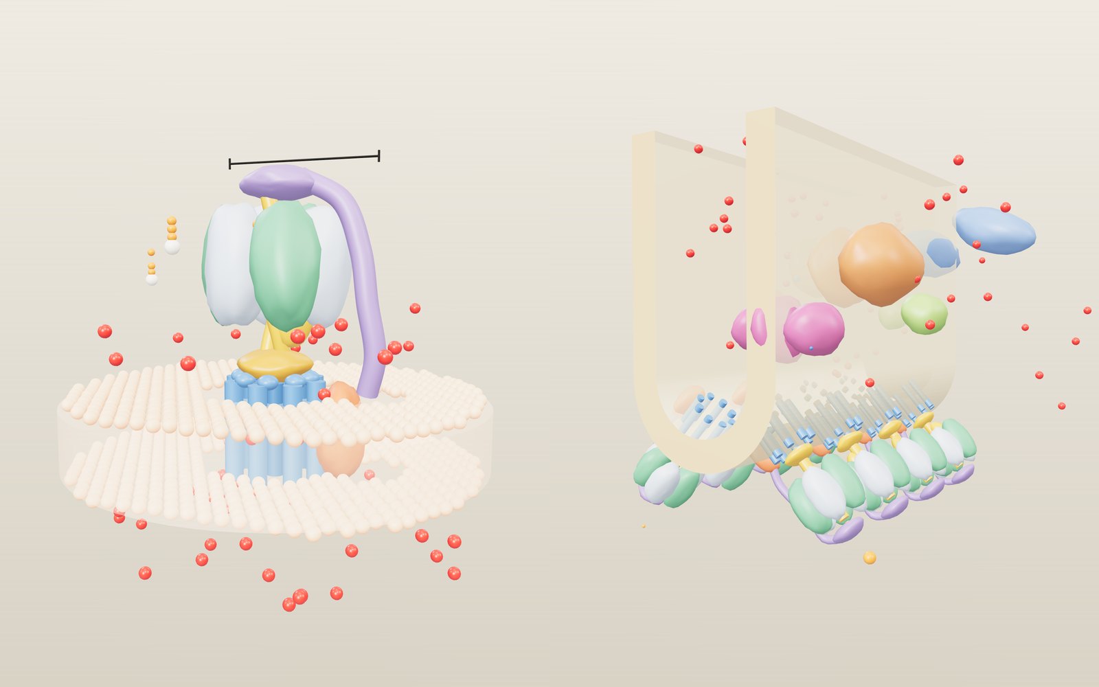
How ATP synthase works
In mitochondria the rotor of ATP synthase turns about 100 times a second, and every full turn releases three molecules of ATP.
Walk inside a cell: animal, plant and bacterium
Grown a million times, a mitochondrion is about the size of a sofa, a whole E. coli is 2 m long, and the kinesin towing a vesicle past you walks at 0.8 metres per second.
How to do CPR, use an AED, help someone choking and use a fire extinguisher
Push 100 to 120 times a minute, about 5 cm deep: with a bystander doing that, survival in Korea in 2024 was 2.36 times higher.
Reaction time test: how many milliseconds do you take?
Light from the screen reaches your eye almost at once, yet your finger moves about a fifth of a second later. Most of that time passes inside your head.