Loading the model
This device cannot show the 3D model. You can read the guide steps below as text.
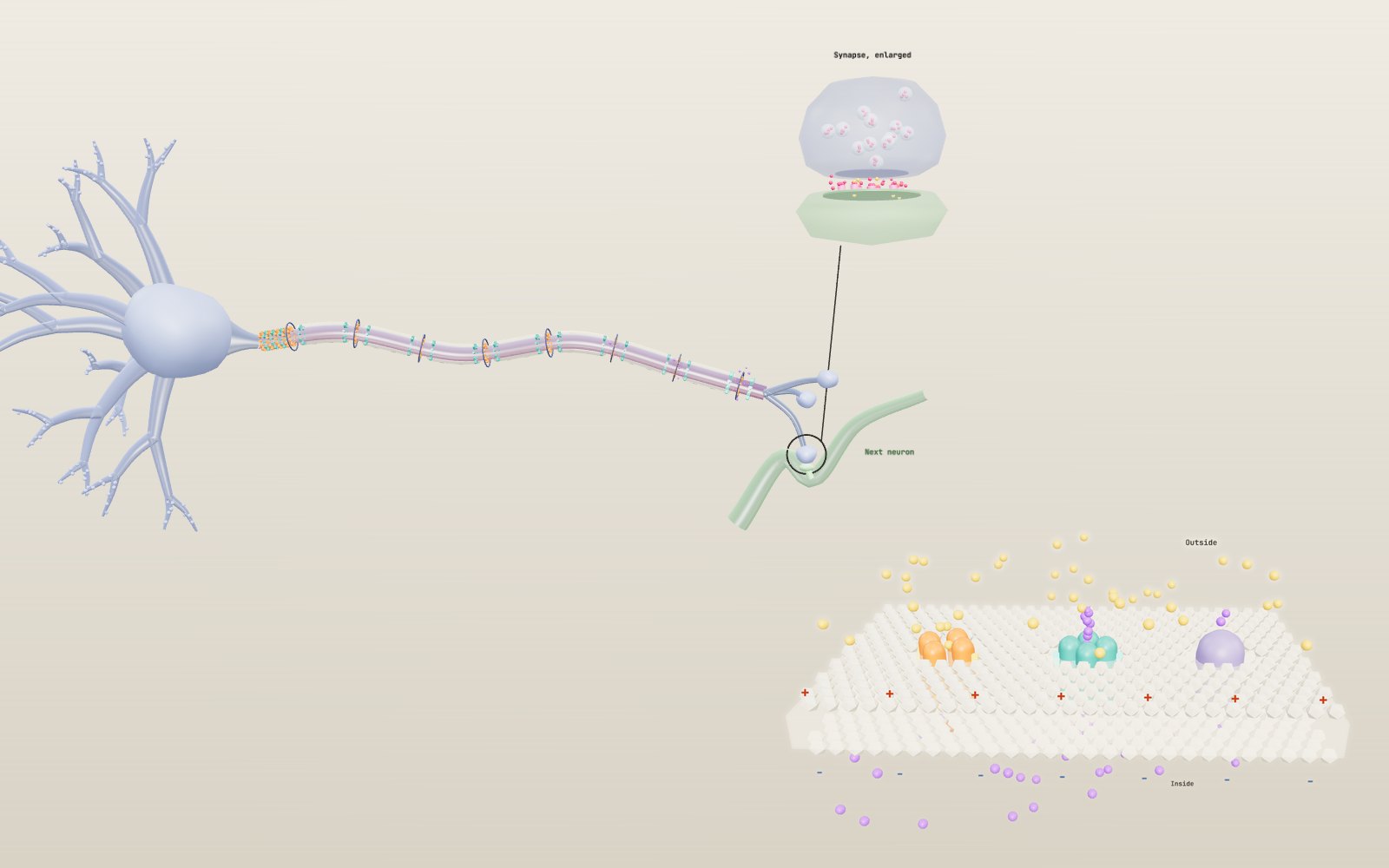
1 / 28 One neuron from end to end Signals come in through the dendrites on the left and leave through the axon terminals on the right. The axon here is shortened and drawn far thicker than life. The panel in the corner plots the membrane potential at the axon hillock. The bar under the model opens seven more scenes.
Drag to turn. Scroll or pinch to zoom.
Guide Parts Controls
01 One neuron from end to end Signals come in through the dendrites on the left and leave through the axon terminals on the right. The axon here is shortened and drawn far thicker than life. The panel in the corner plots the membrane potential at the axon hillock. The bar under the model opens seven more scenes.
02 At rest the inside sits at −70 mV With the stimulus at zero nothing happens and the graph stays flat at −70 mV. Potassium leaks out through open channels and leaves the inside negative. The sodium-potassium pump keeps the two ions where they are.
03 A weak input fades away Now the stimulus is +10 mV. Each pulse runs down the dendrites and lifts the hillock to −60 mV, then dies away. It never reaches −55 mV, so the axon stays dark.
04 Past −55 mV the axon fires At +20 mV the input crosses threshold. Sodium channels at the hillock open, Na⁺ rushes in and the potential shoots to +30 mV. Move the Stimulus slider between 15 and 30: the spike starts sooner but never gets taller.
05 Sodium in, then potassium out Close up on a node with the sheath see-through. Orange sodium channels open first and yellow Na⁺ ions flow in. Then the teal potassium channels open, purple K⁺ flows out and the membrane drops back below rest.
06 Leaping between the gaps With myelin, the spike is rebuilt only at the nodes of Ranvier, which light up one after another. Change Axon diameter in Controls and watch the speed in the corner: a 10 µm myelinated fibre conducts at about 57 m/s.
07 The same axon without myelin Without the sheath every patch of membrane has to fire in turn, and the spike crawls along as a continuous wave. At 10 µm the model drops to about 3 m/s, 18 times slower. On screen the gap is narrowed so you do not wait too long.
08 Across the synapse When the spike reaches a terminal, green calcium ions enter, two docked vesicles fuse and pink transmitter crosses the gap. Receptors on the next neuron glow while it is bound and let ions in. This takes about 1 ms in real life.
09 A patch of membrane under voltage clamp This scene zooms in on a square of membrane with one sodium channel, one potassium channel and the pump. The clamp holds it at −65 mV, then steps to −10 mV for 12 ms. The graph plots the currents the clamp has to supply, as Hodgkin and Huxley recorded them.
10 Sodium shuts itself At +20 mV the sodium channel opens within a fraction of a millisecond and then the red ball on the inside swings into the pore. Sodium current stops even though the voltage stays high. Potassium current keeps flowing as long as the step lasts.
11 The pump trades three for two Back at rest, nothing flows through the voltage-gated channels. The pump keeps working, three Na⁺ out and two K⁺ in for each ATP. It is drawn far slower than a real pump works.
12 A race over one metre Two axons of 10 µm, one wrapped in myelin and one bare, start together. The myelinated signal hops from node to node and covers the metre in 17.5 ms. The bare one needs 316 ms, 18 times longer.
13 Make the bare axon as thick as a squid's Bare axon width is now 476 µm, the size of the axon Hodgkin and Huxley used. It reaches about 22 m/s by being 50 times wider than the myelinated fibre beside it.
14 Damaged myelin Damaged myelin switches on a stretch with no sheath in the middle of the fibre. In the model the signal crawls through it at bare-axon speed. In a real nerve it may stop altogether. This is the idea behind demyelinating diseases such as multiple sclerosis.
15 Glutamate opens channels At a glutamate synapse most receptors are ion channels. They glow while transmitter is bound and the next cell gets a fast excitatory potential of a few milliseconds.
16 Dopamine works more slowly Dopamine binds only receptors that switch on G proteins inside the cell. The response is small, starts later and lasts longer. Its job is to change how the cell answers other inputs.
17 Clearing the cleft Serotonin is pulled back into the terminal by a transporter. Switch Reuptake off and the molecules stay in the cleft longer, and the response stretches out. Some drugs act on this step; this is a concept, not advice.
18 The knee jerk Tap the tendon below the kneecap and a spindle in the thigh muscle fires. The signal runs up a sensory fibre to the spinal cord, crosses one synapse onto a motor neuron and comes back down. The model adds 20 ms for a 175 cm person; measured values are 17 to 21 ms.
19 The ankle jerk takes longer Tap at Ankle sends the signal from the calf, almost twice as far. At the same height the model gives 33.5 ms. Change Height to see why taller people have slower reflexes.
20 Four inputs at once Four excitatory synapses fire together. Each adds 4 mV in the model, and together they reach 16 mV above rest, enough to pass threshold at the trigger zone. This is spatial summation.
21 Spread them out in time The same four inputs now arrive 4 ms apart. Each has partly faded before the next arrives and the peak stays below −55 mV. More inputs or closer spacing is needed: temporal summation.
22 An inhibitory synapse can veto Five inputs would fire the cell, but an inhibitory synapse on the cell body subtracts 8 mV at the same moment. The trigger zone stays quiet. Eccles recorded such inhibitory potentials in spinal motor neurons.
23 Neurons come in many shapes The gallery shows eight types drawn at one size. A Purkinje cell spreads its dendrites in a flat fan crossed by about 175,000 parallel fibres. Neuron type in Controls moves along the row.
24 Hodgkin and Huxley's squid axon A giant axon from a squid lies in seawater with a silver wire threaded inside. The graph shows the potential calculated with the 1952 equations at 6.3 °C, the temperature their rate constants were measured at.
25 Find the threshold Pulse current is 7 µA/cm² for 1 ms, just above the threshold of 6.9 µA/cm². Lower it a little and the potential rises and sinks back. Raise it and the spike starts sooner and grows to about +41 mV, then stops growing.
26 Two pulses close together The second pulse comes 9 ms after the first. At 20 µA/cm² it only produces a small bump, because sodium channels are still inactivated and potassium channels are still open. Raise the current or widen the gap to get a second spike.
27 Steady current, a train of spikes With Steady current on, the axon fires again and again. Stronger current fires faster: about 69 Hz at 10 µA/cm² and 87 Hz at 20 µA/cm². This is how strength turns into frequency.
28 Tetrodotoxin TTX at 100% blocks every sodium channel. The pulse still pushes the membrane up, but nothing regenerates and the wave along the axon disappears. Pufferfish toxin works this way; this is a concept, not advice.
Dendrites Branches that take in signals from other neurons. Each input nudges the membrane potential a little, and the nudges spread toward the cell body. The yellow beads are incoming input. Cell body Holds the nucleus and most of the cell's machinery. Its colour follows the membrane potential: blue at rest, amber when depolarized. Axon hillock The cone where the axon leaves the cell body, with the initial segment just after it. Sodium channels are packed densely here, so this is where the membrane first reaches −55 mV and the action potential starts. Axon The cable that carries the action potential away from the cell body. Its colour shows the membrane potential along its length. The drawing is shortened: real axons run from a fraction of a millimetre to about a metre. Myelin sheath Layers of glial membrane wrapped round the axon: Schwann cells in nerves, oligodendrocytes in the brain and spinal cord. It insulates the axon so current reaches the next gap instead of leaking out. Nodes of Ranvier Gaps about 1 µm long between the sheaths. The action potential is rebuilt only here, so it seems to leap from node to node. Sodium channels Orange. Voltage-gated channels that open when the membrane reaches threshold and let Na⁺ rush in, then close themselves within a fraction of a millisecond. Potassium channels Teal. They open a little later than the sodium channels and let K⁺ out, which brings the membrane back down and briefly below −70 mV. Axon terminals The axon ends in small swellings called boutons. When the action potential arrives, calcium flows in and the bouton releases transmitter onto the next cell. Synaptic vesicles Small bubbles full of neurotransmitter (pink dots). A few wait docked at the membrane; calcium makes them fuse with it and empty into the gap. Receptors Proteins on the next neuron that open when transmitter binds. They glow while bound and let positive ions in, which nudges that neuron toward its own threshold. Lipid bilayer Two sheets of fat molecules, heads out and tails in. Ions cannot cross the oily middle, so they need channel proteins. Na⁺ channel (four domains) One protein folded into four similar domains round a pore. The yellow voltage sensors rise when the inside becomes less negative; that is the m gate of Hodgkin and Huxley. Inactivation gate A loop on the inside that swings into the open pore about a millisecond after it opens and holds it shut. This is the h gate, and it is why the spike cannot run backwards. K⁺ channel Four separate subunits round a pore. Their sensors move more slowly, so potassium flows out after sodium has flowed in: the n gate. Na⁺/K⁺ pump Uses one ATP per cycle to move three Na⁺ out and two K⁺ in. Drawn much slower than a real pump. Myelinated fibre Speed about 5.7 m/s for each micrometre of diameter. Only the nodes fire, so the glow hops. Bare axon Speed grows with the square root of diameter: 1 m/s at 1 µm, about 21 m/s at squid size. Damaged myelin A stretch where the sheath is lost, as in demyelinating disease (concept). Here it conducts like a bare axon. Active zone The patch of presynaptic membrane where vesicles dock. Calcium entering through channels here makes them fuse. Ionotropic receptors Receptors that are themselves ion channels. They open within a millisecond of binding transmitter. Metabotropic receptors Seven-helix receptors that switch on a G protein (the pale ball). Slower, longer lasting and able to change the cell's chemistry. Transporters Proteins that pull transmitter back into the terminal or into glia, ending the signal. Astrocyte A glial cell whose thin processes wrap synapses and take up glutamate. Muscle spindle A stretch sensor inside the muscle. A tap on the tendon stretches it for a moment and it fires. Ia sensory fibre The thick myelinated fibre from the spindle. It runs into the spinal cord through the dorsal root. Dorsal root ganglion Holds the cell bodies of the sensory neurons, off to one side of the path. Motor neuron Receives the sensory fibre directly, one synapse, and sends its own axon back to the same muscle. Inhibitory interneuron A short cell that stops the opposing muscle from contracting while the reflex runs. Quadriceps The thigh muscle that straightens the knee. The calf does the same job for the ankle jerk. Spinal cord Grey matter in a butterfly shape, white matter round it. Dorsal horns up, ventral horns down. Excitatory synapses Each adds a small positive potential that fades as it spreads to the cell body. Inhibitory synapse Placed on the cell body near the trigger zone, where it cancels the most input. Trigger zone Axon initial segment. It fires if the summed potential reaches threshold. Squid giant axon A single fibre about 0.5 mm wide from the mantle of Loligo. Colour shows the computed potential along it. Axial wire A silver wire threaded inside the axon. It makes the whole stretch one patch of membrane, the space clamp the 1952 experiments relied on. Stimulating electrodes Deliver the current pulses set by the sliders. Pyramidal cell Cortex. Triangular cell body, one long apical dendrite with a tuft, basal dendrites round the base, axon down. Purkinje cell Cerebellum. A flat fan of dendrites in one plane, crossed by parallel fibres. Inhibitory. Motor neuron Spinal cord. Big cell body, long dendrites, myelinated axon out to a muscle. Retinal bipolar cell Retina. One process up to the photoreceptors and one down to the ganglion cells. Cerebellar granule cell Cerebellum. Tiny cell with four short claws; its axon rises and splits into parallel fibres. Sensory neuron (DRG) Dorsal root ganglion. One stem from the cell body that splits into a branch to the skin and one to the cord. Chandelier cell Cortex. Inhibitory; rows of boutons on the axon initial segments of pyramidal cells. Medium spiny neuron Striatum. Round dendrites dense with spines. Inhibitory, receives dopamine.