FIG. 38How It Works
How does the eye focus on near and far objects? Try it in 3D
The cornea supplies about 43 of the eye's 60 dioptres, so why does the small lens behind it get all the credit for focusing?
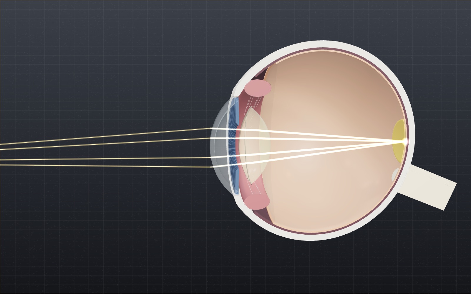
This device cannot show the 3D model. You can read the guide steps below as text.
1 / 11 Two lenses in one eyeLight from the chart bends first at the cornea, about 43 D, then at the lens, about 19 D. Together they make a model eye of 58.8 D, which brings a distant object to a point on the retina 24.2 mm behind the front of the cornea.
Drag to turn. Scroll or pinch to zoom.
Reading a page, then looking out of the window
Look at the words on this screen, then at something across the room. For a moment the far object is soft, and then it snaps into focus. Nothing in your eye moved forward or back the way a camera lens does. A small clear lens behind the pupil changed its shape instead, and it did so in a fraction of a second.
So how does the eye focus on near and far objects? The cornea does most of the bending, about 43 of the eye's 60 dioptres, and the lens adds up to about 11 more at 25 by rounding up, which is enough to bring a page 9 cm away into focus. This exhibit follows that change in a model eye cut in half along its axis. Light from an eye chart is traced through the cornea and the lens to the retina, and every distance and power in the panel comes from the same short calculation. Beside the eye, a window called "What this eye sees" draws the scene the way that eye would see it, and the same picture appears upside down where it really lands, on the retina. You can drag the chart, the lens and a pair of glasses, change the age of the eye, try short sight, long sight, presbyopia and astigmatism, switch to a colour vision deficiency and find the blind spot.
This exhibit explains optics. It is not medical advice. If your vision changes, or something about your eyes worries you, see an eye doctor.
Where the bending happens: cornea and lens
An eye has two lenses in a row. The first is the cornea, the clear dome at the front. In the Exact Gullstrand schematic eye, a standard textbook model, the cornea has a power of 43.05 dioptres. Most of that bending happens at its front surface, where light passes from air (refractive index 1.000) into tissue with an index of about 1.376. Behind the cornea the light crosses the aqueous humour (1.336) and reaches the crystalline lens.
The lens is about 9 mm across and 3.6 mm thick when it is relaxed. Its index rises from about 1.386 in the outer layers to about 1.406 in the core. Because the fluid around it is almost as dense optically, each lens surface bends light only gently, and the whole lens comes to about 19 dioptres in Gullstrand's model. Together the two give the eye roughly 60 dioptres, and the cornea supplies about three quarters of it.
So why does the lens get the credit for focusing? The cornea is fixed. The lens is the only part whose power can change, and that change is what lets the same eye see a mountain and a menu.
Dioptres: the unit for focusing power
Opticians measure a lens by its power in dioptres (D), the reciprocal of its focal length in metres. A lens that brings parallel light to a point 0.5 m away has a power of 1/0.5 = 2 D. Powers add when thin lenses are close together, which is why prescriptions are written in dioptres and not in millimetres.
The same unit describes the light arriving at the eye. Light spreading out from a point 25 cm away has a vergence of −1/0.25 = −4 D. From 6 m it is −0.17 D, close to the zero of truly parallel light from a star. To focus the near point, the eye must find about 4 D more power than it uses for the star. That extra power is called accommodation.
Inside the eye the distances are divided by the refractive index of the fluid. For the model's total power of 58.8 D, the image of a distant object forms 1.336 / 58.8 m = 22.7 mm behind the eye's equivalent lens, which puts the retina of the model eye 24.2 mm behind the front of the cornea. Gullstrand's more detailed eye gives 58.6 D and 24.4 mm.
How the lens changes shape
The lens hangs inside a ring of muscle, the ciliary muscle, on hundreds of fine fibres called zonules. When you look far away the muscle is relaxed and wide, the zonules are pulled tight, and they stretch the lens into a flatter shape. When you look close, the muscle contracts. The ring gets smaller, the zonules go slack, and the elastic capsule around the lens lets it round up.
In Gullstrand's numbers the front surface of the lens goes from a radius of 10 mm to 5.33 mm, the back surface from 6 mm to 5.33 mm, and the thickness from 3.6 mm to 4.0 mm. The front of the lens moves 0.4 mm toward the cornea while the back stays put. The lens power rises from 19.1 D to 33.1 D for 10.9 D of accommodation. Drag the chart toward the eye in the model and you will see the ring of muscle tighten, the fibres fade as they slacken and the lens bulge. Turn on Focus by hand and you can do the bulging yourself: drag the lens up to round it and watch the picture sharpen or blur.
Working out the numbers
The model treats the cornea and lens as two thin lenses 5.4 mm apart in a medium of index 1.336, which is the paraxial (Gaussian) approximation used in most optics textbooks. The combined power is
P = P_cornea + P_lens − (t / n) × P_cornea × P_lens = 43.05 + 19.11 − (0.0054 / 1.336) × 43.05 × 19.11 = 58.8 D.
For an object at distance d, the light reaching the cornea has vergence V = −1/d. The model carries that vergence through the cornea, across the 5.4 mm gap to the lens, adds the lens power and finds where the image forms. It then asks how much the lens must change for that image to sit on the retina.
- 6 m: the eye needs 0.17 D of accommodation. The lens goes to 19.3 D and its thickness barely changes (3.61 mm).
- 25 cm: the eye needs 4.00 D. The lens rounds up to 24.7 D and 3.76 mm thick, and the whole eye reaches 63.5 D.
- 10 cm: the eye needs 10.0 D. The lens reaches 32.6 D and almost 4.0 mm, close to the limit in Gullstrand's table.
How much accommodation is available depends on age. A widely used clinical estimate, Hofstetter's average, is 18.5 − 0.3 × age dioptres. That gives 15.5 D at 10 years (a near point of 6.5 cm), 11 D at 25 (9.1 cm), 6.5 D at 40 (15.4 cm), 2 D at 55 (50 cm) and 0.5 D at 60, when the nearest sharp point has moved out to about 2 m. The age slider in the model uses this formula, and the "Your age" box under the controls turns your own age into an average near point with it.
The pupil: brightness and sharpness
The iris is a ring of muscle in front of the lens. It opens the pupil to about 8 mm in the dark and closes it to about 2 mm in bright light. Light admitted grows with the area of the opening, so an 8 mm pupil lets in (8/2)² = 16 times as much light as a 2 mm one. The readout shows this as "Light in", compared with a 2 mm pupil.
A small pupil also hides focusing errors. When the image forms in the wrong place, each point spreads into a small disc on the retina. The angle that disc covers is roughly the pupil diameter in metres multiplied by the focusing error in dioptres. With a 5 mm pupil and 1 D of error the blur is 0.005 radians, about 17 minutes of arc. For comparison, a letter on the 6/6 line of a chart (20/20 in feet) is 5 minutes of arc tall. That is why people with mild myopia often see better in sunshine than at dusk, and why squinting helps. In the model, the myopic eye at 6 m blurs by 74′ with the room light at 10%, 47′ at 50% and 20′ at 90%.
Myopia: when the image falls in front of the retina
In myopia (short-sightedness) distant things look blurred. The US National Eye Institute describes the usual cause as an eyeball that has grown too long from front to back, though the shape of the cornea or lens can also play a part. The optics are strong enough for a normal eye but too strong for this length, so light from a distant object comes to a point before it reaches the retina and spreads out again.
The myopia setting in the model is an eye that needs −3.00 D glasses. To make that eye, the back of the globe is moved 1.17 mm further from the cornea, which works out to about 2.5 D for each extra millimetre. A chart at 6 m now focuses 1.11 mm in front of the retina. The lens cannot help, because it can only add power and the eye already has too much. Close objects are another matter: anything within about 34.5 cm of the eye is sharp without effort.
A concave (minus) lens fixes this by spreading the light a little before it reaches the cornea. A −3.00 D lens worn 12 mm in front of the cornea has the same effect as −2.90 D at the cornea itself; the difference comes from the gap, and it is why contact lens and spectacle prescriptions differ for stronger corrections. Switch on the glasses and the image moves back onto the retina.
Hyperopia and presbyopia: when the image lands behind
Hyperopia (long-sightedness) is the opposite: an eye too short for its optics, so the image lands behind the retina. A young hyperopic eye can make up the difference by accommodating, even for distant objects. The hyperopia setting needs +4.00 D glasses and is 1.52 mm shorter than the normal model. At 6 m it already uses 4.37 D of its accommodation. At age 40 it has 6.5 D available, and a page at 25 cm needs 8.20 D, so the eye falls 1.70 D short and the image lands 0.56 mm behind the retina. A +4.00 D convex lens adds the missing power; with it, the page at 25 cm needs only 4.40 D, well within reach.
Presbyopia is not a fault in the shape of the eye. With age the lens becomes harder and less flexible, and the National Eye Institute notes that almost everyone notices it, usually after 45. A 55-year-old eye in the model has about 2 D of accommodation, so its nearest sharp point is 50 cm away. Reading at 33 cm needs 3.03 D, and the image lands 0.40 mm behind the retina. Reading glasses of +2.00 D make up the shortfall, leaving 1.13 D for the lens to provide. Look at the far chart through the same glasses and the image moves 0.70 mm in front of the retina, which is why reading glasses blur the room and why many people end up with bifocals or varifocals.
Finding your blind spot
The optic nerve carries the retina's signals to the brain. Where it leaves the eye there are no light receptors, so every eye has a blind spot. Measurements put its centre about 15° to 20° from the centre of vision on the temple side of the visual field, with a width of 5° to 7° and a height of 7.5° to 10°. In the model the optic nerve leaves the retina about 4.5 mm from the centre, which is where a ray arriving at 15° lands.
Switch on the blind spot test and a card appears with a cross and a dot. Close your left eye and look at the cross with your right eye. Move slowly toward the screen or away from it. When your eye is about 3.7 times the gap between the marks away (1 / tan 15° = 3.73), the dot falls on your blind spot and disappears. With marks 6 cm apart that is about 22 cm. You never notice the gap in daily life because the other eye covers it and the brain fills in the missing patch.
What 20/20 and 1.0 actually measure
An eye chart does not measure how sharp a picture is. It measures the smallest detail you can still tell apart, and it states that detail as an angle. On a standard Snellen chart the 20/20 line is built from letters 5 minutes of arc tall (a minute of arc, written 1′, is 1/60 of a degree), with every stroke and every gap in the letter 1′ wide. The American Academy of Ophthalmology puts the meaning in plain terms: with 20/20 vision you can read from 20 feet what an average person reads from 20 feet. The top number is your distance from the chart, and the bottom number is the distance at which a person with typical vision could read the same line. A 20/40 eye must stand at 20 feet to read what the typical eye reads from 40 feet.
The same idea gives the decimal numbers used in Korea, Japan and much of Europe. Decimal acuity is the reciprocal of the smallest detail in minutes of arc, so 1′ is 1.0, 2′ is 0.5 and 0.5′ is 2.0. The metric chart writes 1.0 as 6/6, because 6 m replaces 20 ft. All of these are the same measurement in different dress: 20/20 = 6/6 = 1.0.
Here is what 1′ means in real sizes, using the exhibit's own eye. At 6 m, one minute of arc spans 6000 mm × tan(1′) = 1.75 mm, so a full 20/20 letter is about 8.7 mm tall. The model eye has a total power of 58.8 D, which puts its focal length in air at 1/58.8 = 17.0 mm, and a detail of 1′ therefore makes an image on the retina about 17.0 mm × 0.000291 = 4.9 µm across. That is about two cones wide in the centre of the fovea (see the next section), so 20/20 sits near what a healthy retina can resolve, and only a tiny region of the retina reaches it. This is a rough estimate that treats the eye as a single thin lens, not a measurement of any real person.
Two facts from the AAO keep the number in proportion. A person can have 20/15 vision, which is sharper than average, so 20/20 is not perfect sight. And only about 35% of adults have 20/20 vision without glasses, contact lenses or surgery, while about 75% reach it with correction. In most US states a driver needs 20/40 or better, and "legally blind" means 20/200 or worse with the best correction. Those cutoffs are 2′ and 10′ strokes.
The blur numbers in the pupil section use the same unit. A 20/20 letter is 5′ tall, so the 20′ of blur the myopic eye has at 90% room light is already four letter heights wide, and the 74′ at 10% is about fifteen. That gap is the reason an uncorrected −3 D eye cannot read even the big letters at 6 m.
Rods, cones and why only the centre sees detail
The retina is not an even sheet. In 1990 Christine Curcio and her colleagues at the University of Washington counted photoreceptors in eight whole retinas from seven donors aged 27 to 44. They found an average of 4.6 million cones (range 4.08 to 5.29 million) and 92 million rods (range 77.9 to 107.3 million). So rods outnumber cones by about 20 to 1, and yet detail vision depends on the cones.
The reason is where they sit. Cone density peaks at the centre of the fovea, with an average of 199,000 cones per square millimetre (individuals ranged from 100,000 to 324,000). One millimetre away from the centre the density is lower by an order of magnitude. The very centre of the fovea has no rods at all: the rod-free zone is on average 0.350 mm across, or 1.25°. Rods are densest on an elliptical ring at roughly the eccentricity of the optic disc.
With that density you can check the 20/20 estimate. If cones were packed in a square grid at 199,000 per mm², neighbours would sit about 1/√199000 mm = 2.2 µm apart. A 1′ detail of 4.9 µm therefore covers about two cones from edge to edge, which is the minimum needed to see a gap between two strokes. At 1 mm from the fovea, where cone density is ten times lower, the spacing grows to roughly 7 µm (the square root of ten times 2.2). By the same two-cones rule that is a detail of about 3′, three times coarser. These are rough estimates, since real cones form a hexagonal mosaic and neighbouring cells are pooled. They explain why you point your eyes at a word to read it: fine detail exists only in the small patch that the model labels macula, with the fovea at its centre.
Curcio's counts also show how much one person differs from another. Peak cone density varied by a factor of three between donors, while the total number of cones in the fovea was about the same, which suggests that the cones were packed in differently as the eye developed. For a reader this means that no single "normal" retina exists, and that the model's average numbers describe a typical eye, not yours.
A worked prescription: from diopters to centimetres
Here is how a prescription turns into distances, using the −3.00 D eye of the exhibit. The calculation uses the thin lens rule from the dioptre section and ignores the small details discussed earlier.
- Far point. A −3.00 D lens corrects a relaxed eye when it forms a virtual image at 1/3 m = 33.3 cm, so the far point of the eye is 33.3 cm from the spectacle lens. The glasses sit 12 mm in front of the cornea, which makes it 34.5 cm from the cornea, the same figure quoted above. The focal length of the lens is f = −1/3 m = −33.3 cm.
- Near point at 25. The eye already has 3 D too much power when relaxed. With 11 D of accommodation (Hofstetter, age 25) it can handle light spreading as strongly as 3 + 11 = 14 D, which is a distance of 1/14 m = 7.1 cm from the spectacle plane, about 8.3 cm from the cornea. A young −3 D eye sees clearly between roughly 8 and 34 cm without glasses.
- Near point at 55. With 2 D left the limit is 3 + 2 = 5 D, so the nearest sharp point is 1/5 m = 20 cm (about 21 cm from the cornea). The clear range is now 21 to 34 cm. A book held at 30 cm is comfortable without glasses, which is why a short-sighted person over 50 often takes the glasses off to read.
- Reading glasses on a normal eye. At 55 a normal eye has a near point of 50 cm. A +2.00 D lens in front of it adds 2 D, so to first order the near point becomes 1/(2 + 2) m = 25 cm. The exhibit's own calculation includes the 12 mm gap and finds that at 33 cm the lens needs only 1.13 D of accommodation.
These distances are first estimates. They use one average accommodation formula and ignore the small shifts from lens thickness and from the gap between glasses and eye, so a real person will differ by some centimetres.
Surgery and implants: real changes to the two lenses
Everything above can be changed in a clinic, and the exhibit's numbers show why each procedure works.
LASIK reshapes the cornea. The American Academy of Ophthalmology describes LASIK as using a laser to change the shape of the cornea. The surgeon cuts a thin flap, folds it back, reshapes the tissue underneath with an excimer laser and puts the flap back. To correct myopia the laser removes more tissue from the centre, which flattens the front surface and lowers the cornea's power. How much tissue? An AAO clinical article gives Munnerlyn's rule: depth in micrometres = S² × D / 3, where S is the width of the treated zone in millimetres and D the correction in dioptres. For a 6 mm zone that is 12 µm for every dioptre, so a 4 D correction removes about 48 µm at the centre, and a 3 D correction like the exhibit's eye about 36 µm. The same article notes that larger zones and blended or wavefront treatments remove more tissue.
You can check the result with the keratometer rule, which converts radius of curvature to power with an index of 1.3375 (an assumption built into clinical instruments, which differs from the real tissue index). A 43.25 D cornea has a radius of 337.5/43.25 = 7.80 mm. Lowering it by 3 D to 40.25 D means a radius of 337.5/40.25 = 8.39 mm. A change of 0.59 mm in the radius and a few tens of micrometres of tissue correct an eye that needed −3.00 D glasses. LASIK can leave an eye under- or over-corrected, and the AAO says these are often improved with glasses, contact lenses or a further laser treatment.
Cataract surgery replaces the lens. A cataract is a cloudy lens. The National Eye Institute reports that more than half of Americans aged 80 or older have cataracts or have had surgery for them. In the operation the surgeon removes the clouded natural lens, the 19 D part of the model, and puts in an artificial intraocular lens (IOL). The AAO explains that the surgeon measures the length of the eye and the curve of the cornea, and the results set the IOL's focusing power. The most common type is a monofocal IOL, which has one focusing distance, and most people have it set for distance and wear glasses to read. This is the presbyopia in the model again: the new lens is clear but fixed, so the exhibit's age slider matters even more after surgery. Multifocal and accommodating IOLs exist to widen the range.
Myopia is becoming more common. In 2016 Brien Holden and colleagues published a meta-analysis in the journal Ophthalmology of myopia studies published since 1995. They estimated 163 million people with high myopia in 2000, 2.7% of the world's population. Their projection for 2050 is 4,758 million people with myopia (49.8% of the world) and 938 million with high myopia (9.8%). These are projections with wide confidence ranges (43.4% to 55.7% for myopia in 2050), not measurements. They show why a model of a too-long eye, like the exhibit's, describes a condition that many people will meet.
Astigmatism and colour vision
Astigmatism. In an astigmatic eye the cornea or the lens is curved more steeply in one direction than in the other, so lines in one direction focus at a different depth from lines at right angles to them. The National Eye Institute lists blurry or distorted vision, squinting and eye strain as the common signs and glasses or contact lenses as the usual correction. In the model the Astigmatism slider adds power in one direction only. The view then smears along that direction instead of blurring evenly, and a Glasses cylinder of the same size cancels it.
Colour vision deficiency. Most people have three kinds of cone, peaking at about 420, 534 and 564 nm. The National Eye Institute says the most common colour vision deficiency makes red and green hard to tell apart, that another type makes blue and yellow look alike, and that about 1 in 12 men have some form of it. The Colour vision control uses the filters published by Machado, Oliveira and Fernandes in 2009 for the complete loss of one cone type (protanopia, deuteranopia and tritanopia). Real deficiencies are often milder than these filters. On the dot plate in the view, a 5 drawn in orange-red dots among olive-green dots of similar brightness disappears with the first two filters. The plate is our own drawing, not a clinical test plate.
The ageing lens also turns yellow. The lens absorbs more blue light with age (Pokorny, Smith and Lutze measured this in 1987), so the model tints the view slightly toward yellow as the Age slider rises. The amount is a model value, not a measurement for any one person.
Other eyes
Not every eye focuses the way ours does. An octopus has a camera eye like ours, but its nerve fibres run behind the receptors, so it has no blind spot, and it focuses by moving the whole lens back and forth like a camera (Hanke and Kelber, 2020). A wedge-tailed eagle was measured at 132 to 143 cycles per degree, more than twice the best human value of about 60 (Reymond, 1985; Campbell and Green, 1965). A scallop forms its picture with a curved mirror of guanine crystals instead of a lens (Palmer and others, 2017), and a nautilus has an open pinhole and no lens at all (Muntz and Raj, 1984). A box jellyfish has real lenses, yet its retina sits closer than the sharp image, so it sees a blurred picture on purpose (Nilsson and others, 2005). The "Other eyes" list under the controls has fourteen animals with their sources, and each one links to the How animals see exhibit, where you can look through those eyes.
Common misunderstandings
- "The lens does most of the focusing." The cornea supplies about 43 of 60 D. The lens matters because it can change.
- "20/20 means perfect sight." It means average. Some people see 20/15, and the AAO reports that only about 35% of adults reach 20/20 without glasses, contacts or surgery.
- "The image on the retina is the right way up." It is upside down and reversed left to right, as for any converging lens forming a real image. The brain learns to interpret it, and no part of the eye flips it back.
- "A blind spot is a dark hole in the picture." You do not see black. You see nothing there and the brain fills it in from the surroundings, as the blind spot test shows when a dot vanishes and the card seems unbroken.
- "The pupil sets the focus." The pupil sets how much light enters and how large the blur from any error is, but it does not change the position of the image. Squinting helps for the same reason: a smaller opening reduces blur, as the pupil section computed.
- "Short sight and ageing cancel out." They partly do for reading, and not at all for distance. At 55 a −3 D eye sees clearly between about 21 and 34 cm without glasses, which suits a book, but it still needs glasses to see the road.
Trying it on the screen
The page opens with a short flight from outside a smooth mannequin head, in through the pupil and onto the retina. Press Skip or touch the stage to stop it. After that, each of these takes about a minute.
- See the picture on the retina. Look at the panel above the eye. It is the view in the corner turned upside down, because that is how the cornea and the lens lay it on the retina.
- Focus by hand. Turn on Focus by hand and drag the chart to 25 cm. The view blurs. Drag the lens upward until the letters sharpen, and read the lens power, about 24.7 D. Turn it off and the model focuses for you again.
- Fit glasses. Press Myopia, then drag the glasses from where they rest onto the eye, or switch Glasses on, and change Glasses sphere until the far chart is sharp. The answer for this eye is −3.00 D.
- Near sharp, far soft. Bring the chart close and watch the house behind it blur in the view. At 25 cm a 25-year-old eye blurs the house by about 66′.
- Pupil against blur. Set Eye's refraction to −1.00 D with no glasses and change Room light. A small pupil in bright light makes the same error look much sharper, which is why squinting helps.
- Astigmatism. Set Astigmatism to 1.50 D and watch the view smear in one direction. Put on glasses and raise Glasses cylinder to 1.50 D to straighten it.
- Colour vision. Bring the chart to 50 cm and change Colour vision. With the red or green filter the 5 on the dot plate vanishes, and the red apple and roof lose their contrast with the grass.
- Hide the apple. Turn on Blind spot test and move Gaze to the left until the red apple falls into the dark patch. In real life you would not see a patch: the brain fills it in. The card on the screen helps you find your own blind spot with the 3.7 times rule above.
- Your age. Type your age in the "Your age" box under the controls to see the average near point for that age, then press Use on the stage.
The Challenges list under the controls turns these into eleven tasks, and the stage badge counts how many you have solved. Six faint sparks in the eye open short facts when you tap them.
What the model leaves out
- Thin lenses. Real corneas and lenses have thickness and aspheric surfaces, and the lens has a graded index. The model uses two thin lenses, so its eye is 24.2 mm long instead of Gullstrand's 24.4 mm.
- Axial length only. Myopia and hyperopia are drawn as a longer or shorter eye. In real eyes the cornea and lens shapes also vary.
- The fovea sits on the axis. In a real eye the line of sight is about 5° away from the optical axis.
- Average accommodation. Hofstetter's formula is an average; individuals vary by several dioptres, and the minimum expected value is lower.
- Lens shape. The lens outline uses Gullstrand's surface radii. Because those are spheres, the drawn lens narrows more at its edge than a real lens does when it rounds up.
- Distances not to scale. The chart moves between 35 mm and 85 mm in front of the drawing. The ray calculation uses the true distance.
- The view is a model. The blur in "What this eye sees" is the model's blur circle for the chart and for a house 6 m away, drawn as an even blur. Real blur also depends on the eye's other optical errors, and the brain sharpens what it sees.
- Astigmatism in one direction. The model adds the extra power along one fixed direction and lets the eye focus for the other, so the view smears in that direction only.
- Colour filters at full strength. The colour vision filters simulate the complete loss of one cone type. Many people with a colour vision deficiency have a milder, shifted cone instead.
- The blind spot outline. The dark patch is an outline about 5.5° wide and 7.5° tall, 15.5° from the centre of gaze. Its real size and shape vary from person to person.
A short history
Allvar Gullstrand, a Swedish ophthalmologist, received the 1911 Nobel Prize in Physiology or Medicine "for his work on the dioptrics of the eye". His schematic eye, with surfaces, thicknesses and indices for both relaxed and focused states, is still printed in optics textbooks and is the source of the main numbers in this model. The age rule in the model is younger. In 1950 Hofstetter fitted straight lines to Duane's earlier measurements of focusing range at different ages, giving an average, a minimum and a maximum amplitude for any age. Eye clinics still use these lines to judge whether a patient focuses as well as expected, and the model takes the average one.
Questions
Is the lens or the cornea more important for focusing?
The cornea does more of the bending, about 43 of roughly 60 dioptres. The lens matters because its power can change. Without that change you could see clearly at only one distance.
What does a prescription like −2.50 mean?
It is the power of the spectacle lens in dioptres. A minus sign means a concave lens for myopia, a plus sign a convex lens for hyperopia or reading. A −2.50 eye sees sharply without glasses out to about 40 cm.
Why do I need reading glasses after 40 or so?
The lens stiffens with age, so the ciliary muscle can no longer make it round enough. The near point moves out from about 10 cm in your twenties to 50 cm or more in your fifties. Reading glasses add the power the lens can no longer supply.
Why can't I see my blind spot with both eyes open?
Each eye's blind spot falls on a part of the scene the other eye can see. Even with one eye closed, the brain fills the gap with the surrounding pattern.
Why do things look sharper in bright light?
A bright scene makes the pupil smaller. A smaller opening shrinks the blur disc made by any focusing error, so slight myopia or hyperopia matters less.
Does looking at screens for a long time make the lens tired?
Holding focus at a near distance keeps the ciliary muscle contracted, and many people feel that as strain. The model shows how much accommodation a distance needs. Whether screen use changes your eyes over time is a medical question; ask an eye doctor.
Can an eye be both myopic and presbyopic?
Yes. Presbyopia affects the range of focus, myopia the distance at which the eye is sharpest when relaxed. A myopic person over 50 may read comfortably without glasses but need them for driving.
What does 20/20 mean, and is it the best vision?
It means you read at 20 feet what an average person reads at 20 feet. It is not the best: 20/15 is sharper than average, and the smallest detail a 20/20 eye resolves is a stroke of 1 minute of arc.
Why do I see best when I look straight at something?
The centre of the fovea holds the most cones, about 199,000 per square millimetre on average, and has no rods. A millimetre away the cone density is ten times lower, so fine detail needs the eye to point at it.
Does LASIK change the lens?
No. LASIK reshapes the cornea with a laser. For a 6 mm treated zone it removes about 12 micrometres of tissue per dioptre of myopia, so −3 D takes about 36 µm. The natural lens stays, and it still stiffens with age.
What happens to focusing after cataract surgery?
The cloudy lens is replaced with an artificial lens whose power is chosen from the length of the eye and the curve of the cornea. The most common type has one focusing distance, so reading glasses are usually needed for near work.
Is myopia becoming more common?
A 2016 meta-analysis projected that about half of the world's people (49.8%) will be myopic by 2050, up from a far smaller share in 2000. It is a projection with a wide confidence range.
Why does the image on the retina appear upside down?
A converging lens forms a real image that is inverted. The retina records it that way and the brain interprets the signals as an upright scene.
Terms
- Dioptre (D): the power of a lens, equal to 1 divided by its focal length in metres.
- Vergence: how fast light is spreading or converging, in dioptres. Light from a point d metres away has a vergence of −1/d.
- Accommodation: the extra focusing power the lens adds for near objects.
- Amplitude of accommodation: the most accommodation an eye can produce, which falls with age.
- Near point: the closest distance at which an eye can focus.
- Far point: the farthest distance an eye can focus without accommodating. It is infinitely far for a normal eye.
- Myopia: an eye that focuses distant objects in front of the retina.
- Hyperopia: an eye that focuses distant objects behind the retina when relaxed.
- Presbyopia: loss of accommodation with age.
- Schematic eye: a set of surfaces, distances and indices that behaves like an average human eye.
Sources
Addresses checked on 2026-10-16 (first sixteen), 2026-10-22 (next eight) and 2026-10-27 (last twelve).
Kaschke, Donnerhacke and Rill, Optical Devices in Ophthalmology and Optometry, chapter 2, "Optics of the Human Eye" (Wiley-VCH sample chapter; Exact Gullstrand eye, table 2.1). https://application.wiley-vch.de/books/sample/3527410686_c02.pdf
HyperPhysics, Georgia State University, Scale model of human eye (cornea, lens, aqueous and vitreous indices). http://hyperphysics.phy-astr.gsu.edu/hbase/vision/eyescal.html
HyperPhysics, Georgia State University, Accommodation (ciliary muscle and fibres, pupil 2 to 8 mm). http://hyperphysics.phy-astr.gsu.edu/hbase/vision/accom.html
HyperPhysics, Georgia State University, The Eye. http://hyperphysics.phy-astr.gsu.edu/hbase/vision/eye.html
OpenStax, College Physics 2e, 26.1 Physics of the Eye. https://openstax.org/books/college-physics-2e/pages/26-1-physics-of-the-eye
OpenStax, College Physics 2e, 26.2 Vision Correction. https://openstax.org/books/college-physics-2e/pages/26-2-vision-correction
National Library of Medicine, StatPearls, Accommodative Insufficiency (total power 58 to 60 D, Hofstetter formulas, D = 1/F). https://www.ncbi.nlm.nih.gov/books/NBK587363/
Marcos and others, The Optical Design of the Human Eye: a Critical Review, Journal of Optometry (PubMed Central). https://pmc.ncbi.nlm.nih.gov/articles/PMC3972707/
National Library of Medicine, StatPearls, Accommodative Excess (Hofstetter's average, minimum and maximum, from Duane's data). https://www.ncbi.nlm.nih.gov/books/NBK592379/
Evidence of the Amplitude of Accommodation of School-Going Children in the 21st Century (PubMed Central; Hofstetter 1950). https://pmc.ncbi.nlm.nih.gov/articles/PMC10289054/
Fine-scale measurement of the blind spot borders (PubMed Central; position 15° to 20°, size). https://pmc.ncbi.nlm.nih.gov/articles/PMC10494866/
National Eye Institute, How the Eyes Work. https://www.nei.nih.gov/learn-about-eye-health/healthy-vision/how-eyes-work
National Eye Institute, Nearsightedness (Myopia). https://www.nei.nih.gov/learn-about-eye-health/eye-conditions-and-diseases/nearsightedness-myopia
National Eye Institute, Farsightedness (Hyperopia). https://www.nei.nih.gov/learn-about-eye-health/eye-conditions-and-diseases/farsightedness-hyperopia
National Eye Institute, Presbyopia. https://www.nei.nih.gov/learn-about-eye-health/eye-conditions-and-diseases/presbyopia
The Nobel Prize, The Nobel Prize in Physiology or Medicine 1911. https://www.nobelprize.org/prizes/medicine/1911/summary/
American Academy of Ophthalmology, What Does 20/20 Vision Mean? (Snellen fractions, 20/15, 35% and 75% of adults, 20/40 and 20/200 limits). https://www.aao.org/eye-health/tips-prevention/what-does-20-20-vision-mean
Curcio, Sloan, Kalina and Hendrickson, Human photoreceptor topography, Journal of Comparative Neurology 292, 1990 (cone and rod numbers, foveal density, rod-free zone). https://pubmed.ncbi.nlm.nih.gov/2324310/
American Academy of Ophthalmology, LASIK: Laser Eye Surgery (reshaping the cornea, flap, under- and over-correction). https://www.aao.org/eye-health/treatments/lasik
American Academy of Ophthalmology, Surface Ablation: PRK, LASEK, Epi-LASIK (Munnerlyn formula, 12 µm per dioptre at 6 mm). https://www.aao.org/education/current-insight/surface-ablation-photorefractive-keratectomy-lasek
American Academy of Ophthalmology, Intraocular Lenses (IOL) for Cataract Surgery. https://www.aao.org/eye-health/diseases/cataracts-iol-implants
American Academy of Ophthalmology, What Are Cataracts?. https://www.aao.org/eye-health/diseases/what-are-cataracts
National Eye Institute, Cataracts (more than half of Americans aged 80 or older). https://www.nei.nih.gov/learn-about-eye-health/eye-conditions-and-diseases/cataracts
Holden and others, Global Prevalence of Myopia and High Myopia and Temporal Trends from 2000 through 2050, Ophthalmology 123, 2016. https://pubmed.ncbi.nlm.nih.gov/26875007/
National Eye Institute, Color Blindness (red-green and blue-yellow types, about 1 in 12 men). https://www.nei.nih.gov/learn-about-eye-health/eye-conditions-and-diseases/color-blindness
National Eye Institute, Astigmatism. https://www.nei.nih.gov/learn-about-eye-health/eye-conditions-and-diseases/astigmatism
Machado, Oliveira and Fernandes, A Physiologically-based Model for Simulation of Color Vision Deficiency, IEEE Transactions on Visualization and Computer Graphics 15, 2009. https://doi.org/10.1109/TVCG.2009.113
Pokorny, Smith and Lutze, Aging of the human lens, Applied Optics 26, 1987. https://doi.org/10.1364/AO.26.001437
Augusteyn, On the growth and internal structure of the human lens, Experimental Eye Research 90, 2010. https://doi.org/10.1016/j.exer.2010.01.013
Bowmaker and Dartnall, Visual pigments of rods and cones in a human retina, Journal of Physiology 298, 1980. https://doi.org/10.1113/jphysiol.1980.sp013097
Campbell and Green, Optical and retinal factors affecting visual resolution, Journal of Physiology 181, 1965. https://doi.org/10.1113/jphysiol.1965.sp007784
Hanke and Kelber, The Eye of the Common Octopus (Octopus vulgaris), Frontiers in Physiology 10, 2020. https://doi.org/10.3389/fphys.2019.01637
Reymond, Spatial visual acuity of the eagle Aquila audax, Vision Research 25, 1985. https://doi.org/10.1016/0042-6989(85)90226-3
Palmer and others, The image-forming mirror in the eye of the scallop, Science 358, 2017. https://doi.org/10.1126/science.aam9506
Muntz and Raj, On the visual system of Nautilus pompilius, Journal of Experimental Biology 109, 1984. https://doi.org/10.1242/jeb.109.1.253
Nilsson and others, Advanced optics in a jellyfish eye, Nature 435, 2005. https://doi.org/10.1038/nature03484
More exhibits
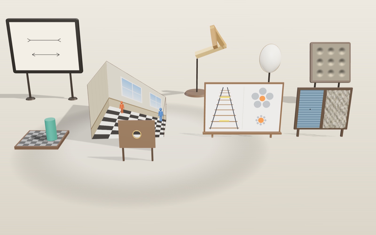
Optical illusions: why equal grey tiles look different
Tile A looks dark and tile B looks light, yet your screen sends the same grey to your eye from both.
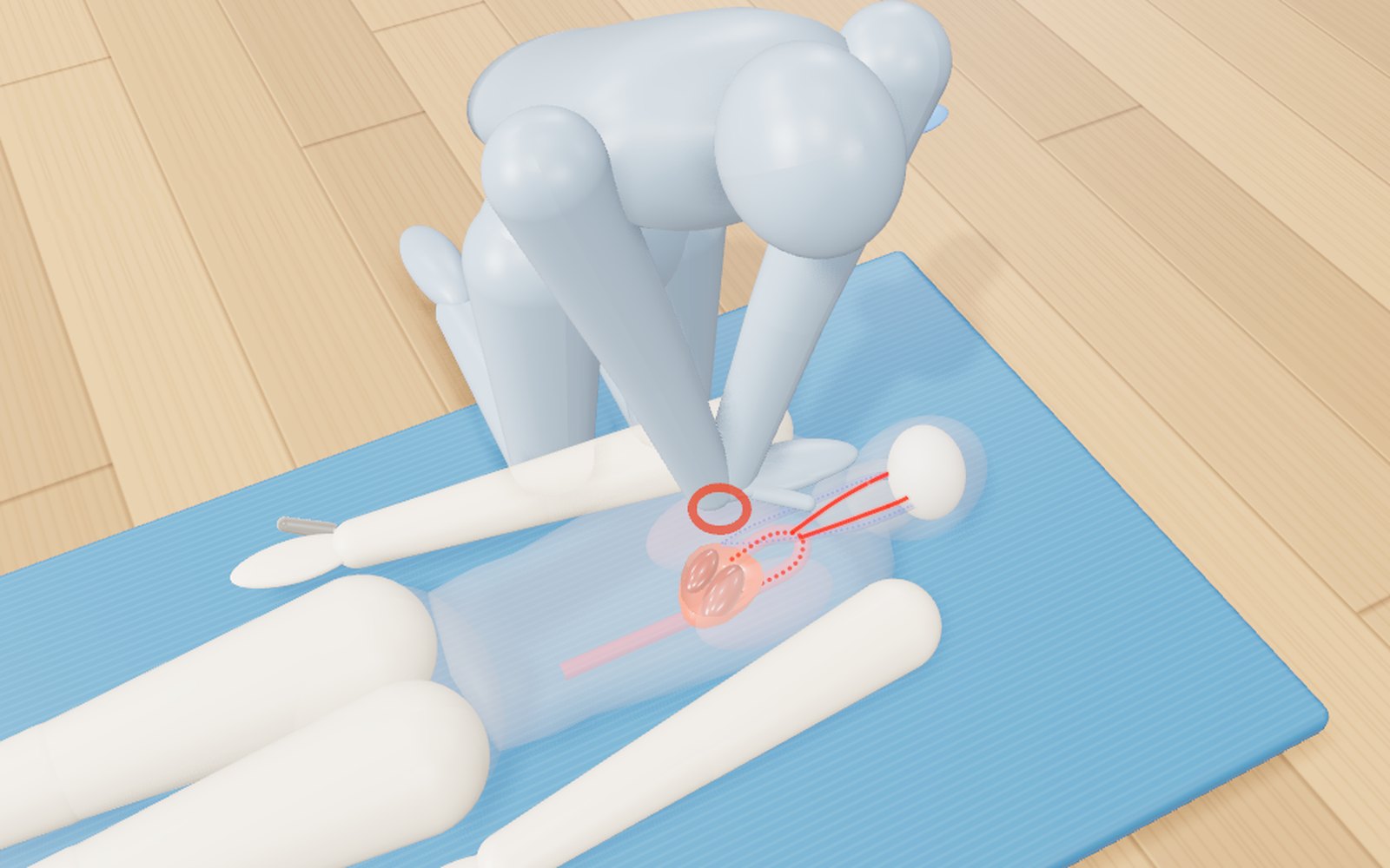
How to do CPR, use an AED, help someone choking and use a fire extinguisher
Push 100 to 120 times a minute, about 5 cm deep: with a bystander doing that, survival in Korea in 2024 was 2.36 times higher.
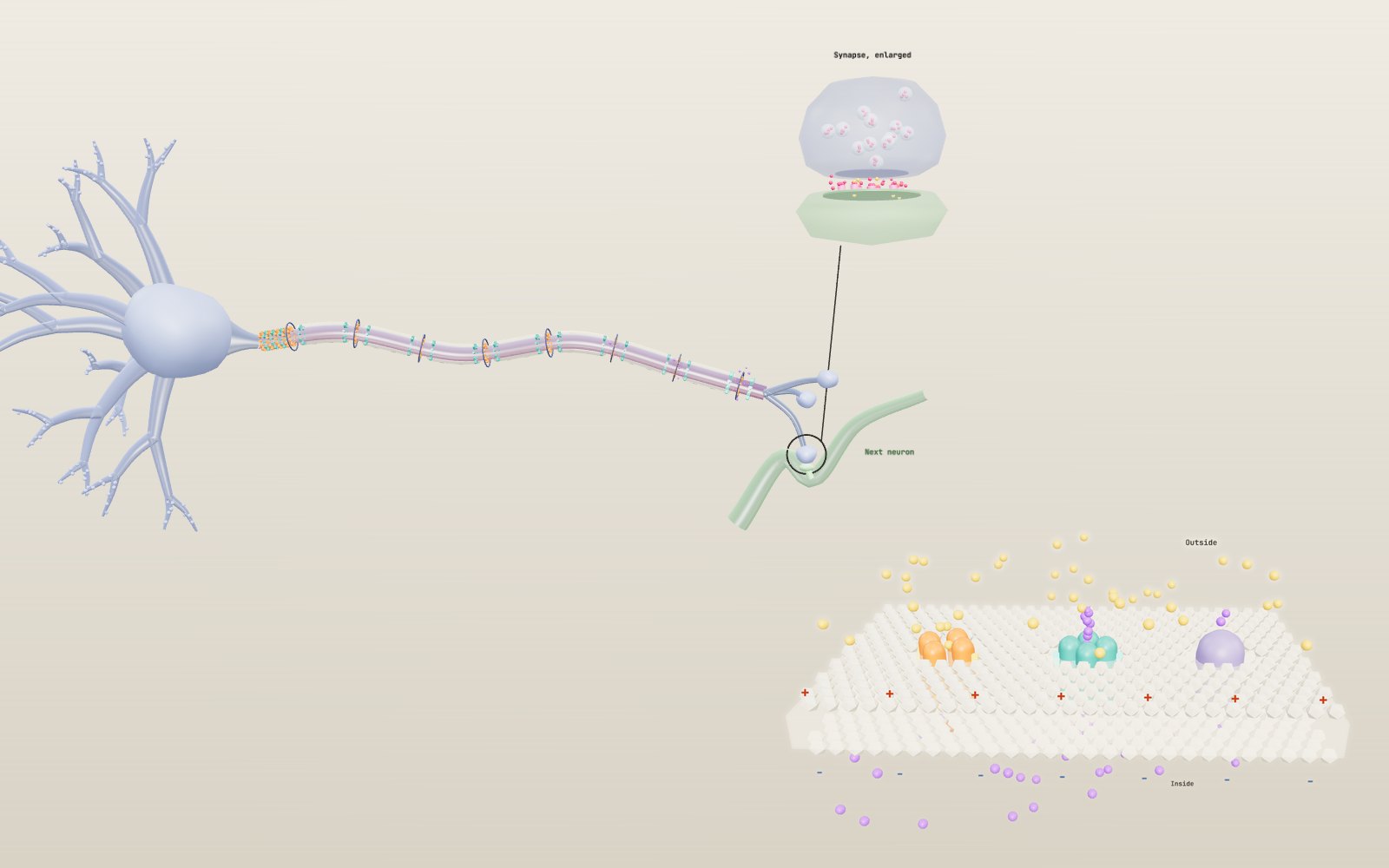
How nerve signals travel
A nerve cell sits at about −70 mV, and when a stimulus lifts it to about −55 mV it fires a spike that is always the same size.
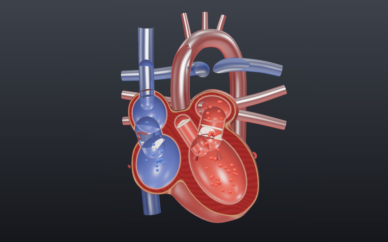
Heart beating: interactive model of the heart, step by step
Your heart pushes out about 70 mL of blood with each beat, and at a resting pace it does that about 108,000 times a day.
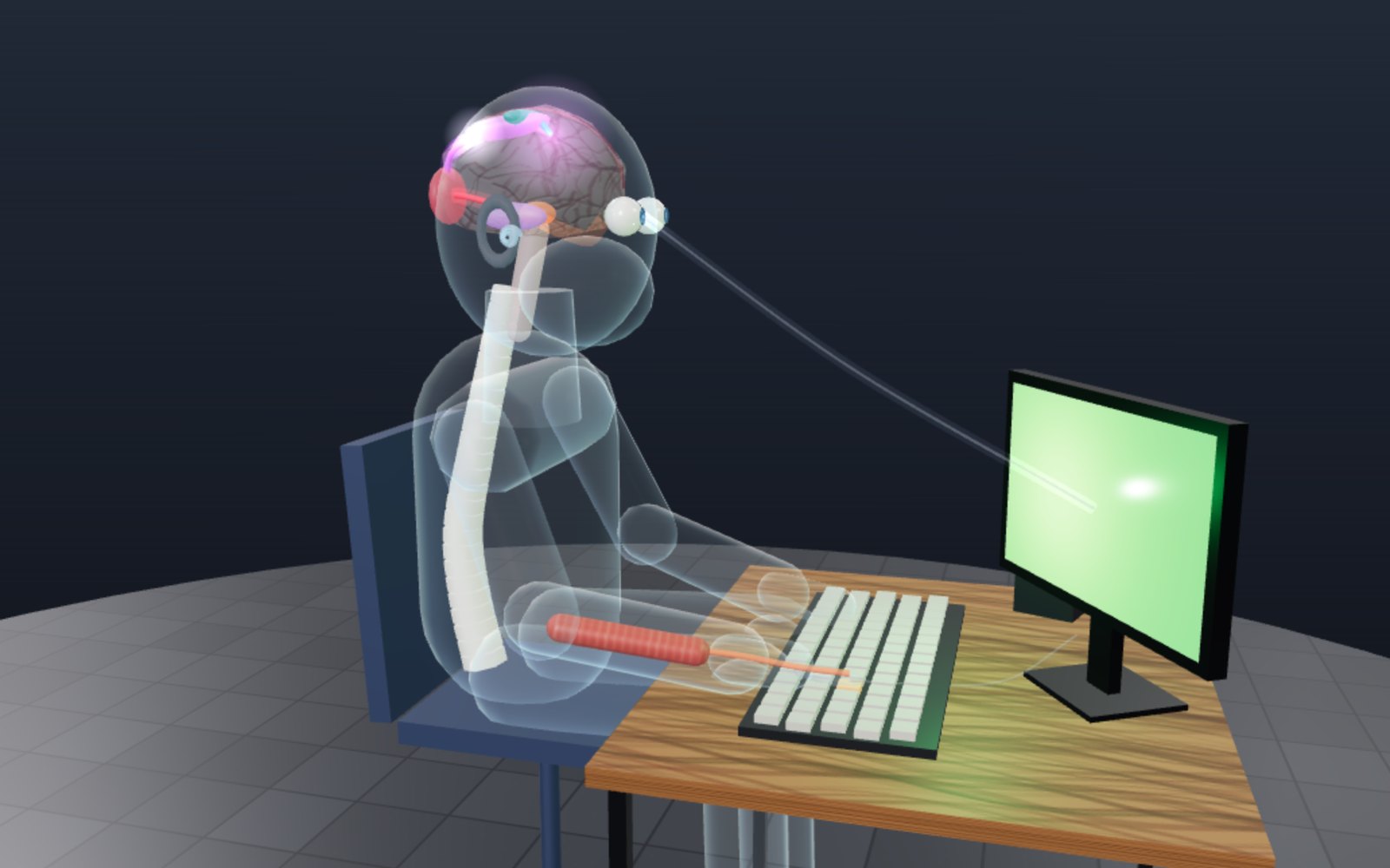
Reaction time test: how many milliseconds do you take?
Light from the screen reaches your eye almost at once, yet your finger moves about a fifth of a second later. Most of that time passes inside your head.
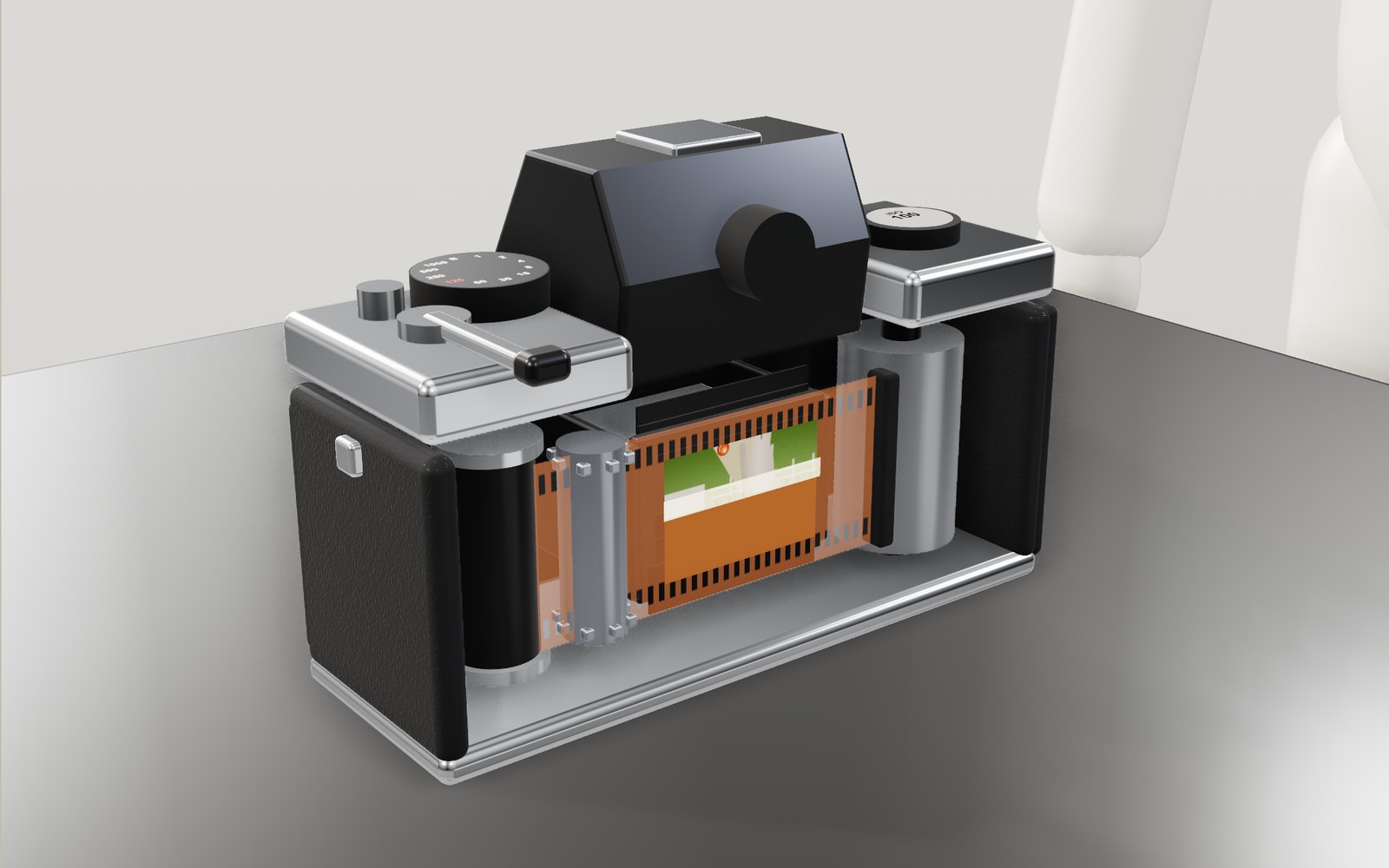
How film cameras and instant cameras work
At 1/1000 of a second a camera shutter never uncovers the whole frame at once: a narrow slit sweeps across the film instead.