Loading the model
This device cannot show the 3D model. You can read the guide steps below as text.
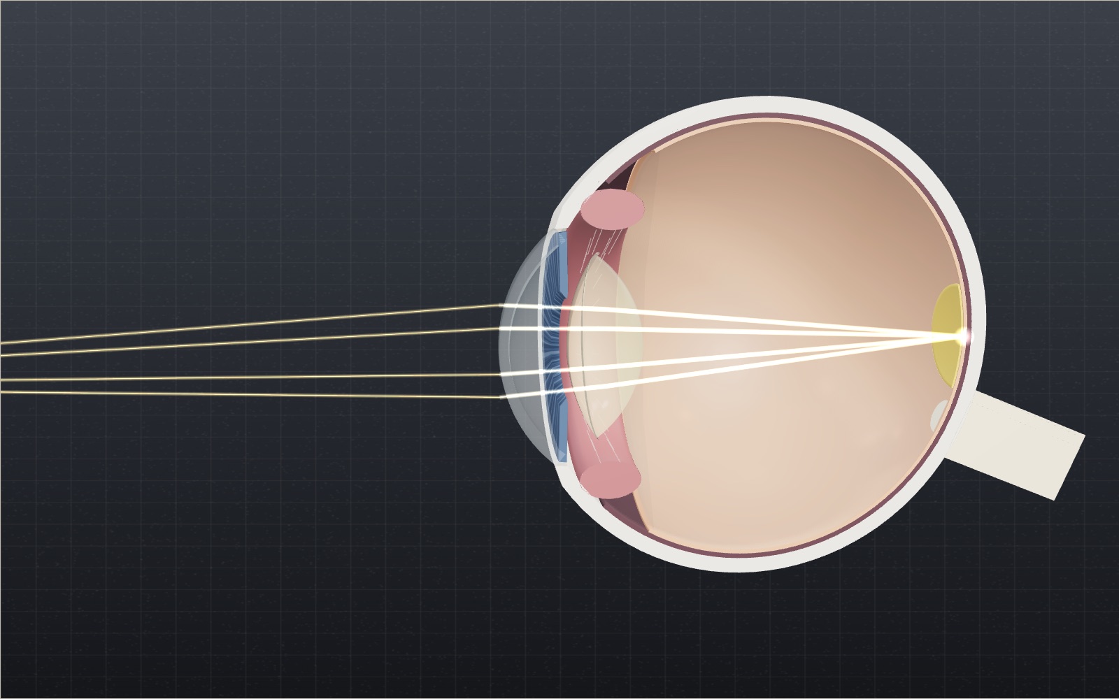
1 / 11 Two lenses in one eye Light from the chart bends first at the cornea, about 43 D, then at the lens, about 19 D. Together they make a model eye of 58.8 D, which brings a distant object to a point on the retina 24.2 mm behind the front of the cornea.
Drag to turn. Scroll or pinch to zoom.
Guide Parts Controls
01 Two lenses in one eye Light from the chart bends first at the cornea, about 43 D, then at the lens, about 19 D. Together they make a model eye of 58.8 D, which brings a distant object to a point on the retina 24.2 mm behind the front of the cornea.
02 Looking at something close At 25 cm the light arrives spreading out, and the eye needs 4 D more. The ciliary muscle tightens, the lens rounds up from 19.1 to 24.7 D and its thickness grows from 3.60 to 3.76 mm. Drag the distance slider toward 10 cm and watch it bulge further.
03 The pupil in dim light In a dark room the iris opens the pupil to about 8 mm. That lets in about 16 times as much light as a 2 mm pupil, but a wide pupil also makes any focusing error look blurrier. Try the room light slider.
04 Myopia: the image falls short This eye is a little too long for its optics. A chart 6 m away comes to a point 1.11 mm in front of the retina, and the letters in the readout turn to fog. Looking closer helps: the clear range ends at about 34 cm.
05 A concave lens moves it back A −3.00 D spectacle lens spreads the light slightly before it reaches the cornea. The image moves back onto the retina and the blur disappears.
06 Hyperopia: the image lands behind This eye is too short, so it must focus even for distant things. At 40 it can add 6.5 D, but a page at 25 cm needs 8.2 D and the image lands 0.56 mm behind the retina. Switch on the glasses to add +4.00 D.
07 Presbyopia and reading glasses At 55 the lens can add only about 2 D, so the nearest clear point has moved out to 50 cm. Reading at 33 cm needs about 3 D. These +2.00 D reading glasses make up the difference. Take them off to see the image slip behind the retina.
08 The blind spot The optic nerve leaves the retina a little toward the nose, and there are no light receptors where it does. Light from about 15° out on the temple side of what you are looking at lands right there, along the blue path. The card on the screen helps you find your own.
09 What this eye sees The window in the corner draws the scene as this eye would see it, and the panel above the eye shows the same picture upside down, the way it lands on the retina. Focused on the chart at 25 cm, the house 6 m behind it blurs by about 66′.
10 Astigmatism Here the eye has 1.50 D of extra power in one direction only, so the view smears that way instead of blurring evenly. Glasses with a 1.50 D cylinder cancel it: try Glasses cylinder in Controls.
11 Colour vision This filter removes the green cones (deuteranopia, after Machado and others, 2009). The 5 on the dot plate vanishes and the red apple blends into the grass. About 1 in 12 men have some colour vision deficiency, according to the National Eye Institute.
Eye chart The object being looked at. The slider moves it from 10 cm to 6 m; the drawing squeezes that range so it fits. Spectacle lens Sits 12 mm in front of the cornea. Concave for myopia, convex for hyperopia and for reading glasses. Cornea The clear front window, about 0.5 mm thick. Its curved front surface does most of the bending, about 43 D. Iris and pupil A ring of muscle that opens the pupil to about 8 mm in the dark and closes it to about 2 mm in bright light. Crystalline lens About 19 D when relaxed. It rounds up and thickens from 3.6 mm toward 4.0 mm when you look at something close. Ciliary muscle and zonules A ring of muscle joined to the lens by fine fibres. When it tightens, the fibres go slack and the lens bulges. Vitreous humour Clear gel filling the back of the eye. Its refractive index, 1.336, is close to that of water. Retina The light-sensitive lining at the back. A sharp image needs the light to come to a point right on this layer. Macula A yellowish patch about 5.5 mm across with the fovea at its centre, where detail vision is sharpest. Optic nerve Carries signals to the brain. Where it leaves the retina there are no light receptors, which makes the blind spot. Sclera and choroid The tough white outer coat, and inside it the dark choroid that carries the blood supply for the retina.