Loading the model
This device cannot show the 3D model. You can read the guide steps below as text.
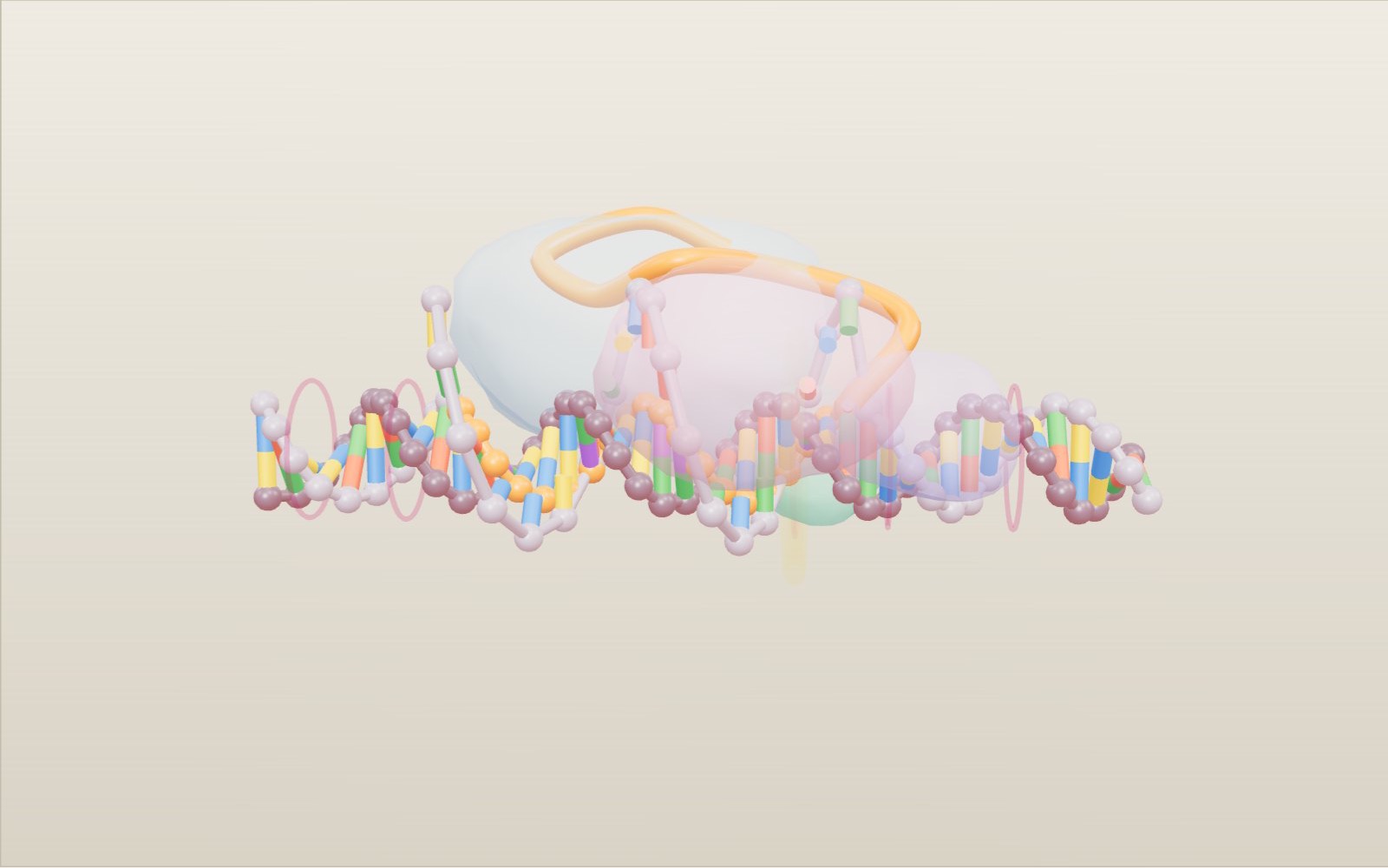
1 / 8 A stretch of DNA, to scale This is 44 base pairs of DNA, about 15 nm long. The black bars give the scale: the helix is 2 nm wide and turns once every 3.4 nm. The sequence is an example made up for the model.
Drag to turn. Scroll or pinch to zoom.
Guide Parts Controls
01 A stretch of DNA, to scale This is 44 base pairs of DNA, about 15 nm long. The black bars give the scale: the helix is 2 nm wide and turns once every 3.4 nm. The sequence is an example made up for the model.
02 Where the guide comes from Bacteria keep pieces of virus DNA between short repeats in their own genome, the CRISPR array. Each piece is copied into RNA that leads a Cas protein to the same virus if it comes back.
03 Cas9 carries the guide Cas9 arrives holding the guide RNA (orange). The protein is drawn see-through so you can follow the RNA. Its 20-letter tip is the part you can change in the box below the model.
04 First it finds a PAM Cas9 grips the DNA only next to NGG. Here the PAM is TGG. Switch PAM (NGG) off in Controls and the protein drifts past without opening the helix.
05 The guide pairs, starting at the PAM The DNA opens one base pair at a time from the PAM end, and the guide pairs with the dark strand while the grey strand is pushed aside. Type a different letter into the guide and the pairing stops at that letter.
06 Two domains, two cuts With all 20 bases paired, the HNH domain (green) swings in and cuts the dark strand. RuvC (pink) cuts the grey one. Both cuts fall 3 base pairs before the PAM.
07 Repair by joining the ends Without a template the cell glues the ends back together. This often adds or loses a few bases; the model shows one extra base pair, marked by the ring.
08 Repair from a template With Repair template on, a matching piece of DNA lies next to the break and the cell copies it. Three base pairs at the cut change colour to the template’s letters.
DNA double helix Two sugar-phosphate strands wound round each other, 2 nm across, one turn every 3.4 nm (10 base pairs). The grey strand carries the same letters as the guide; the dark strand is the one the guide pairs with. Base pairs A pairs with T and C pairs with G, one pair every 0.34 nm. Colours: A green, T red, G yellow, C blue, and U (in RNA) purple. Target (protospacer) The 20 base pairs that match the guide. The guide pairs with the dark strand here and the grey strand is pushed aside into a loop, the R-loop. PAM (NGG) Three bases right after the target on the grey strand: any base, then two Gs. Here it is TGG. Cas9 looks for this first and opens the DNA only next to it. Guide RNA One RNA strand. The first 20 letters (the spacer) choose the target; the rest (the scaffold) holds the guide inside Cas9. It joins the crRNA and tracrRNA that the bacterium uses. REC lobe The recognition lobe of Cas9. It holds the guide RNA and the RNA-DNA pair. Shown as a soft blob, not the measured surface. RuvC domain One of the two cutting domains, in the nuclease lobe. It cuts the grey strand, the one that is not paired with the guide. HNH domain The other cutting domain. It swings into place only when the guide has paired along the target, then cuts the dark strand, the one paired with the guide. PAM-interacting domain The part of Cas9 that reads the PAM. Two arginines recognise the GG pair by hydrogen bonds. Cut site Both strands are cut 3 base pairs before the PAM, at the same place, so the ends are mostly blunt. After the cut, the ring marks the base pairs that the repair changed. Repair template A piece of DNA that matches both sides of the cut and carries three changed base pairs. With it present, the cell can copy the change into the cut site. CRISPR array In the bacterium, short repeats (grey) alternate with spacers (coloured) copied from viruses that attacked before. Each spacer becomes a guide.