Loading the model
This device cannot show the 3D model. You can read the guide steps below as text.
1 / 8 A fist-sized pump A typical adult heart is about 12 cm long and weighs 250 to 350 g. It beats here at 75 times a minute, and every beat the chambers shrink as they empty and swell as they refill.
Drag to turn. Scroll or pinch to zoom.
Guide Parts Controls
01 A fist-sized pump A typical adult heart is about 12 cm long and weighs 250 to 350 g. It beats here at 75 times a minute, and every beat the chambers shrink as they empty and swell as they refill.
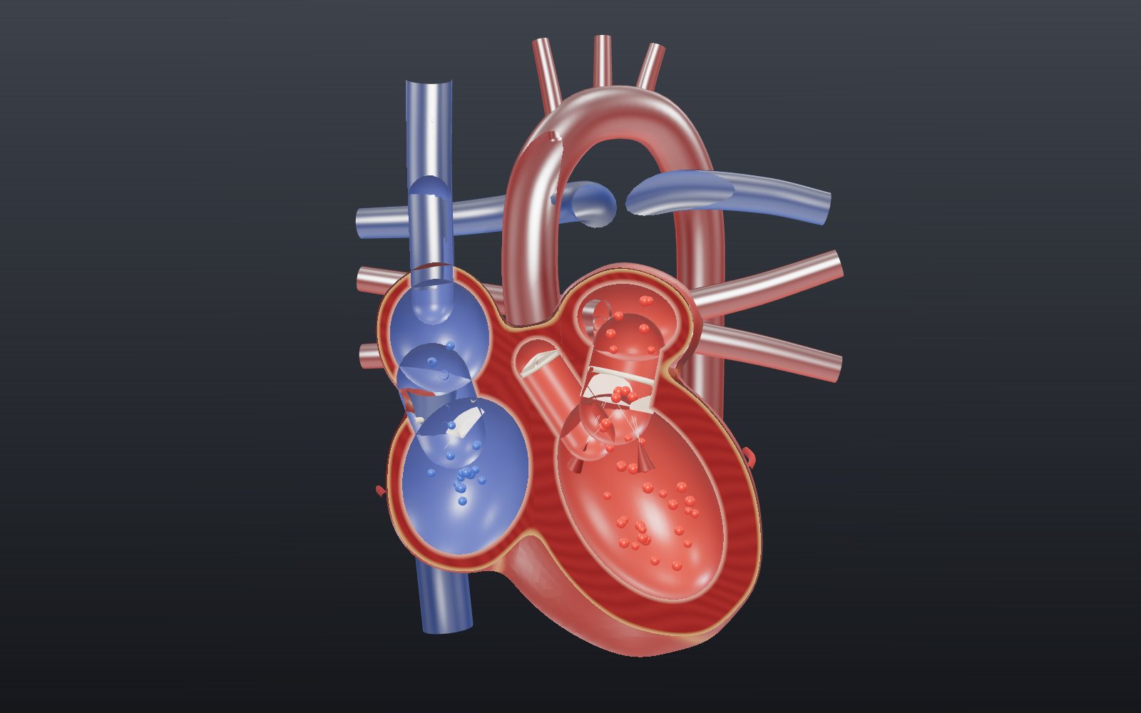
02 Two pumps side by side Cut the heart down the middle and it turns into two pumps. The right side, drawn blue, takes oxygen-poor blood from the body to the lungs. The left side, drawn red, takes oxygen-rich blood from the lungs to the body.
03 Valves that only open one way In slow motion, the tricuspid and mitral valves swing open while the ventricles fill. As soon as the ventricles squeeze, the backflow slaps them shut, and the cords below stop them flipping into the atria.
04 The exits to the arteries The pulmonary and aortic valves open only while the ventricles are pushing. They sit higher up than the cut, so the outer wall is made see-through here. Their three cusps fill like pockets and close when the push ends.
05 The SA node starts every beat A small patch of muscle in the wall of the right atrium fires by itself, about 100 times a minute if nothing slows it. The signal spreads over both atria, which glow yellow as they are triggered, and they squeeze.
06 A pause, then a fast spread The atria and ventricles are insulated from each other except at the AV node, which holds the signal for about 100 ms so the atria can finish pumping. Then the bundle of His and the Purkinje fibres carry it to the apex and up the walls in about 100 ms more.
07 The ECG traces the same events The small P wave in the readout is the atria being triggered, the tall QRS spike is the ventricles, and the T wave is the ventricles resetting. The first heart sound comes just after the R peak, the second near the end of T.
08 Faster beats, more blood per minute Cardiac output is heart rate times stroke volume. At rest that is 75 × 70 mL = 5.25 L a minute. Here the heart runs at 120 beats with 90 mL each, which comes to 10.8 L. Try the sliders yourself.
Right atrium Collects oxygen-poor blood from the body through the two venae cavae. The SA node sits in its wall near the top. Right ventricle Pumps blood a short way to the lungs. The lungs need little pressure, so its wall is much thinner than the left ventricle's. Left atrium Receives oxygen-rich blood from the four pulmonary veins. It sits at the back of the heart. Left ventricle Pumps blood to the whole body against much higher resistance, so its wall is the thickest in the heart. It forms the apex. Tricuspid valve Three flaps between the right atrium and ventricle. Cords tie them to small muscles so they cannot flip back into the atrium. Mitral valve Two flaps between the left atrium and ventricle. Its closing, together with the tricuspid, makes the first heart sound. Pulmonary valve Three pocket-shaped cusps at the exit of the right ventricle. It lies in front of the cut, so use See inside to find it. Aortic valve Three cusps at the exit of the left ventricle. Its closing, with the pulmonary valve, makes the second heart sound. Venae cavae The superior vena cava brings blood from the head and arms, the inferior one from the rest of the body. Pulmonary trunk and arteries Carry oxygen-poor blood to the lungs. They are the only arteries after birth whose blood is low in oxygen. Pulmonary veins Four veins, two from each lung, that bring oxygen-rich blood back to the left atrium. Aorta The main artery. It rises from the left ventricle, arches over the top of the heart with three branches and runs down the spine. Conduction system The SA node, AV node, bundle of His, bundle branches and Purkinje fibres. Specialised muscle cells that set and spread each beat. Papillary muscles and cords Small muscles on the ventricle walls, three on the right and two on the left, tied to the AV valve flaps by the chordae tendineae. Coronary vessels The heart's own blood supply. The coronary arteries leave the aorta just above the aortic valve and run in the grooves between the chambers. Blood (drawn as beads) Blue beads stand for oxygen-poor blood and red beads for oxygen-rich blood. Real blood is dark red or bright red.