FIG. 21Senses
Animal vision: see the world through 69 animals’ eyes
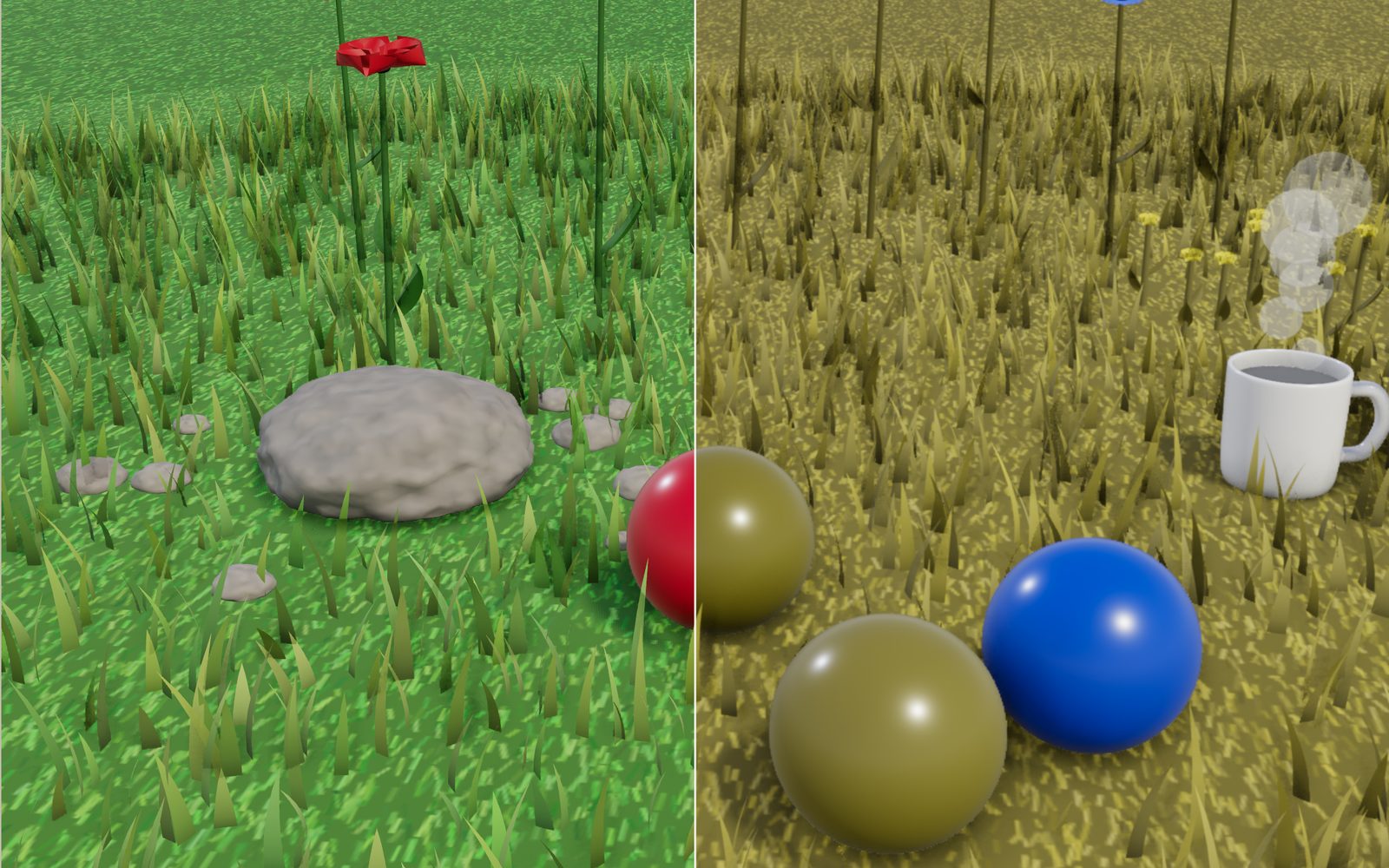
A red ball on green grass is easy for you to find and surprisingly hard for a dog. A honeybee sees a pattern in the same flower that you cannot.
This device cannot show the 3D model. You can read the guide steps below as text.
1 / 20 One garden, 69 pairs of eyesThe same garden is redrawn for each animal. Colour, sharpness, the light an eye needs and how fast it sees all change. Pick an animal in the bar under the picture, or a scene at the top.
Drag to turn. Scroll or pinch to zoom.
Why a dog loses the red ball
Throw a red ball onto a green lawn and you can follow it all the way. Many dogs run straight past it and then find it with their nose. The ball has not become invisible. To a dog, red and green fall on the same kind of cone cell, so the ball and the grass come out as two shades of one dull olive. Throw a blue ball instead and the dog sees it at once.
That small scene holds most of what this exhibit is about. An eye is not a camera that records the world as it is. It samples light with a handful of receptor types, at a certain sharpness, at a certain speed, over a certain angle, and only above a certain brightness. Every animal has settled each of those differently. The stage redraws one garden, a night forest, a reef and a meadow seen from the air through the eyes of 69 animals, and every number it shows comes from a measurement in a paper or a university page listed at the end. Where nobody has measured something, the stage says "no figure" instead of guessing.
How an eye turns light into colour
The retina has two kinds of light sensors. Rods work in dim light and do not tell colours apart. Cones work in daylight and come in a few types, each tuned to a band of wavelengths. No single cone knows what colour it sees: it only reports how much light it caught. The brain compares the reports from different cone types, and that comparison is what we experience as colour.
People usually have three cone types. Measured one cell at a time in a human retina, their peaks sit near 420 nm (violet-blue), 534 nm (green) and 564 nm (yellow-green). The green and the long cone overlap a lot, but a red and a green object still give them different ratios, so we see two colours. Two lights that give all three cones the same catches look identical to us even when their spectra differ, and that is why a screen can fake almost any colour with only three narrow primaries.
The shape of a receptor's sensitivity curve is much the same from pigment to pigment once you know where it peaks. Govardovskii and colleagues fitted a template in 2000 that predicts the whole curve from the peak alone, including a small second bump at short wavelengths. The curves in the stage readout are drawn with that template, one per receptor type, and the colour model uses the same curves.
Two cones: dogs, cats, horses and the farm
Most mammals have two cone types: one for short wavelengths and one for middle-to-long ones. With two cones the eye can still tell blue from yellow, but red, orange, yellow and green all fall mostly on the long cone and differ mainly in brightness. A person with red-green colour blindness sees in a similar way.
In dogs, tests on trained animals published in 1989 found two cone pigments near 429 nm and 555 nm. Cats have the same pair, near 450 and 556 nm, measured in retinal recordings in 1969 and 1979. Horses have theirs at 428 and 539 nm, measured in 2001. Cattle, goats and sheep were tested together in 1998: a short cone between 444 and 455 nm and a long one between 552 and 555 nm. White-tailed deer have a short cone near 450 to 460 nm and a middle one near 537 nm.
Pick the dog and turn on "Half and half". The left half is your view, the right half the dog's. The red and green balls, more than 100 units apart for you on the CIELAB colour-difference scale, come out about 5 units apart for the dog. The blue ball stays more than 100 units away from the grass. If you buy a toy for a dog that plays on grass, blue or yellow is a better choice than red. Try the horse, the cow and the deer too: their long cones sit at slightly different wavelengths, so the reds turn olive by slightly different amounts.
Ultraviolet: mice, bees, budgerigars and goldfish
Many animals see a band of light we cannot: ultraviolet, below about 400 nm. Our own lens absorbs most of it before it reaches the retina. In 1991 Jacobs and colleagues found that some rodents have a cone most sensitive to ultraviolet, the first such receptor known in a mammal. Mice have one peaking at 360 nm and a green one at 508 nm, and most of their cones carry both pigments at once. Rats also have an ultraviolet and a middle-wave cone and, in tests published in 2001, used them together to tell colours apart.
Honeybees have ultraviolet, blue and green receptors, peaking at 344, 436 and 544 nm, and no receptor for red. Budgerigars and zebra finches have four cone types, one of them a true ultraviolet cone at 360 to 380 nm. Goldfish have four as well, at 356, 447, 537 and 623 nm. A flower bat from Central and South America, Pallas's long-tongued bat, responds to ultraviolet down to 310 nm, but in its case one pigment does both jobs: the green pigment's small second absorption band catches the ultraviolet, so the bat sees ultraviolet as brightness, not as a separate colour.
Ultraviolet patterns are common on flowers. Black-eyed Susans look plain yellow to us, but their petals absorb ultraviolet near the centre and reflect it at the tips, which makes a dark ring around the middle of the flower. In a 2014 field study, wild bees visited flower heads whose ultraviolet ring had been enlarged more often than ones whose ring had been made smaller. In the garden, pick the honeybee, the mouse or the budgerigar and look at the yellow flowers close up. Your screen cannot emit ultraviolet, so the stage carries a separate ultraviolet value for every surface and shows it in false colour; with "Ultraviolet only" you see that channel alone.
More receptors is not always better colour
The Japanese yellow swallowtail butterfly has eight varieties of receptor in six classes. In behavioural tests published in 2008, butterflies trained to one colour could tell it from another only 1 nm away at three places in the spectrum, near 430, 480 and 560 nm. That pattern is the mark of four receptor types working together, so the swallowtail sees with four channels even though it has more receptor types than that.
Mantis shrimp are the extreme case. Some species have 12 receptor types spread from 300 to 720 nm. One might expect them to tell colours apart better than anything else alive. Thoen and colleagues tested that in 2014 and found the opposite: the shrimp confused lights 12 to 25 nm apart, coarser than the butterfly and coarser than us. Their suggestion is that the shrimp do not compare receptors finely but recognise a colour by which narrow receptor responds, scanning the eye across an object. The stage models that idea with a simple rule: the strongest of 12 receptors names the colour, so the reef breaks into flat bands of hue. The same animals can also see circularly polarised light, which Chiou and colleagues showed in 2008.
One cone: sharks, octopuses and cuttlefish
Hart and colleagues measured the visual pigments of 17 shark species in 2011. Every one had a single cone type, peaking between 532 and 561 nm. With one cone an animal can judge brightness but not colour, so sharks may well be colour-blind. Whales, dolphins and seals also have a single cone type; they reached the same design on their own.
The common octopus has one visual pigment, peaking at 475 nm, and most behavioural tests find it colour-blind. Cuttlefish are similar, with one receptor type near 500 nm, yet they change the colour of their skin to match the background. Marshall and Messenger called this "colour-blind camouflage" in 1996. What cephalopods have instead is polarisation vision: their receptors sit at right angles to each other, and each absorbs light polarised in one direction best. Under water, where light reflected off silvery fish is strongly polarised, that is a useful signal. Pick the octopus at the reef and turn on "Polarisation": strongly polarised light shows yellow and unpolarised light blue.
How sharp: cycles per degree
Sharpness is measured with stripes. Show an animal pairs of black and white stripes and make them finer until it can no longer tell them from plain grey. The finest pattern it still sees, counted as stripe pairs (cycles) per degree of view, is its acuity. A person with 20/20 vision resolves about 30 cycles per degree; with the optics of the eye bypassed by interference fringes, people resolved gratings of about 60 cycles per degree in a 1965 study. The stage uses 60 as the person's limit and converts any figure into a Snellen fraction with 20/(600 ÷ cycles per degree).
Dogs reach 11.6 to 12.6 cycles per degree in recordings from brain and retina, about a fifth of the best human figure. Horses reached 23.3 at best in behavioural tests. Cats are given as 3.5 to 9, mice about 0.5 and rats about 1. A tawny owl tested in bright light beside two people saw less detail than they did and about as much as a pigeon. The wedge-tailed eagle is at the other end: 132 to 143 cycles per degree in Reymond's 1985 tests, more than twice the human figure and close to the 157 its pupil can let through.
Insects build their eyes from many small facets, each looking in its own direction. The angle between neighbouring facets sets how much detail they can pick up. It ranges from tens of degrees in primitive insects to 0.24 degrees in dragonflies, the finest known. The front of a honeybee's eye resolves about 0.54 cycles per degree. The principal eyes of the jumping spider Portia are tiny telephoto cameras with receptors 2.4 minutes of arc apart, about 12.5 cycles per degree, much better than any insect of its size. Scallops, with up to 200 small eyes along the edge of the shell, resolve about 2 degrees.
To show acuity on a screen, the stage blurs the picture by an amount that depends on how many pixels one degree of the view covers. A grating of c cycles per degree needs two pixels per cycle, so a view with p pixels per degree can show at most p ÷ 2 cycles per degree. In the garden one degree is only about 16 to 20 pixels tall on a 1440 px wide window and about 8 to 10 on a phone held upright, which is enough for 4 to 10 cycles per degree. That is coarser than a dog's eye, so at the normal view the dog's blur works out to a third of a pixel and the dog looks as sharp as you. The picture is not wrong: the screen itself is coarser than both eyes.
"Look far" fixes that the way a telescope would. It narrows the camera's view by 2, 4 or 8 times, so the same degree is spread over 2, 4 or 8 times as many pixels, and the blur is drawn at its true size for that view. At ×4 on a laptop one degree covers about 70 to 80 pixels: your half stays sharp, the dog's half blurs by about 2.5 to 3 pixels and the honeybee sees only a few facets. On a phone use ×8, where one degree covers about 70 pixels. The readout shows the pixels per degree and the blur at the current magnification. An eye sharper than yours, such as the eagle's, cannot be shown at all: the picture still has to pass through your own eyes, so the stage leaves it unblurred and says so.
The sky scene does the same with distance. It shows a meadow through a view only 4 m tall, like a telescope, at a distance you choose from 20 m to 1.5 km. At about 480 m a 40 cm rabbit spans about 3 cycles for you and about 7 for the eagle: a blob for you, still a rabbit for the eagle. Two rabbits sit 2.4 m apart so that with "Half and half" each side of the picture has one.
How fast: flicker fusion
Shine a light that switches on and off and raise the rate. At some frequency the flashes merge into a steady glow. That frequency is the critical flicker fusion frequency, and it says how quickly an eye can follow change. Inger and colleagues collected measurements for 81 species in 2014. People sit at about 60 Hz, cats at 55, dogs at 80, pigeons between 77 and 143 depending on the study, chickens between 72 and 105, and blowflies, honeybees and dragonflies at 200 to 300. Sharks measured so far sit between 18 and 37 Hz.
The lantern in the garden and the lamp post in the forest flicker at the rate you set, from 20 to 320 Hz. A screen cannot show a light flickering 100 times a second, so the lantern pulses slowly when the animal you picked could see its flicker, and the pulse grows deeper the further the lantern's rate is below the animal's fusion frequency. At 100 Hz, the flicker of a lamp on a 50 Hz mains supply, the lantern is steady for you and pulses for a bee. At 60 Hz, the refresh rate of many screens, it stays steady for a cat and pulses for a dog.
The numbers side by side
The atlas under the controls lists all 69 animals with their sources. Here are the ones with the most measurements, in the units the stage uses. A dash means no figure was found in these sources.
| Animal | Receptor peaks (nm) | Acuity (cpd) | Field | Flicker fusion (Hz) |
|---|---|---|---|---|
| Person | 420, 534, 564 | 60 | 180° | 60 |
| Dog | 429, 555 | 11.6 to 12.6 | 240° | 80 |
| Cat | 450, 556 | 3.5 to 9 | 200° | 55 |
| Horse | 428, 539 | 23.3 | 350° | - |
| Mouse | 360, 508 | 0.5 | - | - |
| Rat | ultraviolet and middle-wave | 1 | - | 30 |
| Honeybee | 344, 436, 544 | 0.54 | - | 200 to 300 |
| Pigeon | 409, 453, 507, 567 | - | - | 77 to 143 |
| Goldfish | 356, 447, 537, 623 | - | - | - |
| Wedge-tailed eagle | violet-sensitive short cone | 132 to 143 | - | - |
| Tawny owl | - | - | 201° | - |
| Dragonfly | 15 to 33 opsin genes | 2.1 | - | 300 |
| Jumping spider | ultraviolet and green | 12.5 | - | - |
| Sharks | one cone, 532 to 561 | - | - | 18 to 37 |
Read across a row and you can see the trade-offs. The eagle has the sharpest eyes but no special night ability on record; the cat sees in dim light but blurs fine detail; the bee sees ultraviolet and fast flicker but resolves only about half a cycle per degree.
Seeing at night
In dim light the cones stop working and the rods take over. Rods are more sensitive, but there is one kind, so colour fades away. People lose colour at about 0.02 cd/m², a level around twilight. Cats see in dimmer light than we do: tested with the same patterns, they were 0.74 log units, about 5.5 times, more sensitive, and most of that came from their larger pupils and the light-gathering of their optics. Barn owls have tubular eyes with an f-number of 1.13; a person's eye in the dark is about f/2.4, so the owl's image on the retina is about 4.5 times brighter. The stage uses that ratio as the barn owl's night gain and labels it as a model.
Two animals keep colour where we cannot. Helmet geckos have no rods at all, only three kinds of large cone, and in 2004 they told blue from grey at 0.002 cd/m², dim moonlight. Elephant hawkmoths chose coloured targets at 0.0001 cd/m², dim starlight, in tests published in 2002; in the same light people are completely colour-blind. Set the night forest to "Dim starlight" and try them. For these two the stage brightens the picture by the ratio of their colour limit to ours (10 and 200 times), and marks that as a model too.
Many night-active animals have a mirror behind the retina, the tapetum lucidum. Light that passes the receptors without being caught is sent back through them for a second chance. Dogs, cats, horses, cattle and many other mammals have one, and so do sharks. It is why their eyes shine back in a torch beam. Turn on the torch in the forest and look at the deer.
How wide: fields of view
Eyes on the front of the head see the same scene twice, which helps judge depth; eyes on the sides see more of the world at once. A person sees about 90 degrees to each side, about 180 degrees together, with about 120 seen by both eyes. A dog's field is about 240 degrees with 30 to 60 degrees of overlap, a cat's about 200. A horse covers about 350 degrees and is blind only straight behind and right in front of its forehead. Each eye of a tawny owl covers only 124 degrees, the narrowest field measured in any vertebrate, so its total field is 201 degrees with 48 degrees of overlap. Chameleons move their two eyes independently while they search, then lock both onto prey.
The panorama shows the whole circle around the camera, with your forward direction in the middle. The animal's field covers the middle of the strip; whatever lies outside it is hatched. For animals with no field figure the panorama shows all 360 degrees and says so.
Pupil shapes
Pupils come in round, slit, bar, W and pinhole shapes. Banks and colleagues compared 214 land animals in 2015 and found a pattern. Grazing prey animals such as goats, sheep, horses and deer have horizontal pupils, and their eyes roll as the head tips down to graze so the pupil stays parallel to the ground. That gives a sharp, wide view along the horizon. Small predators that ambush from close to the ground, such as cats, tend to have vertical slits, which help judge distance by blur. Cuttlefish have W-shaped pupils that even out light coming from above and below. The pupil of a helmet gecko closes to a row of pinholes.
The pupil sets the shape of the blur for anything out of focus. Turn on "Pupil blur" and pick the goat: objects in front of and behind the point the camera turns around smear sideways, while vertical edges stay sharper. The cat's slit does the opposite. The blur strength here is a model chosen so the shape shows clearly; the shape is the animal's.
Under water
Water absorbs light, and red goes first. Pope and Fry measured pure water in 1997: at 650 nm it absorbs 0.34 per metre, at 450 nm only 0.0092, about 37 times less. In the reef scene the light travels from the surface down to the reef and then across to your eye, and each colour is weakened along that path. At 15 m only about 2 per cent of the red is left, so the red fish turns grey-brown even for you, while blue and yellow fish keep their colour longer. Real sea water also scatters light and carries plankton; the stage uses pure water and a simple haze.
Heat: pit vipers
Pit vipers, pythons and boas have pits on the face that detect infrared from warm bodies. Bullock and Cowles recorded from the nerves of the pit in 1952 and showed that it responds to warmth nearby. In 2010 Gracheva and colleagues found the molecule behind it: the nerve fibres supplying the pits carry TRPA1 channels, which open when warmed. Infrared warms the pit membrane, and the channels report the change. It is a sense of heat, not a third kind of eye. The stage paints warm surfaces in false colour over a dim view: the tea glows, the sun-warmed stone glows faintly, and the open sky is cold.
The eye lab
The fifth scene puts the eyes themselves on a table. A human eye cut in half shows the cornea and lens bending light to a point on the retina, the image upside down, with the fovea where the cones are packed most densely. A compound eye is a dome of hexagonal facets; part of it is cut away to show the long ommatidia, each with its own lens and receptors. A third eye has a tapetum behind the retina, with a ray going in and coming back out. Five irises show the pupil shapes. The last model is a scallop eye, which focuses with a curved mirror instead of a lens. Palmer and colleagues showed in 2017 that the mirror is tiled with square guanine crystals and sends light onto two retinas, one for the centre of the view and one for the edge. Pick an animal while you are in the lab and the camera moves to its eye type.
Sixty-nine animals and the eyes they share
Version three of this exhibit more than doubles the cast, from 31 to 69 animals in ten groups. The new ones were chosen because people ask about them: farm animals and pets, zoo animals, birds of the garden and the shore, reptiles and frogs, the insects that bite or pollinate, and a few sea creatures whose eyes are odd enough to be worth a look. Each was added only with a paper that measured something about its eye, and the number on the stage links to that paper.
Some animals have not been measured directly but clearly share an eye system with one that has. The atlas marks them "grouped with" another animal, and the reason for grouping is itself a source. A chimpanzee is grouped with a person, because studies of primate photopigments find that Old World monkeys, apes and people all have three cone types with nearly the same peaks. A seal is grouped with the bottlenose dolphin: antibody staining found no blue cones in any of the five seal and sea lion species or the seven toothed whales examined, while their land relatives kept them. Pythons and boas are grouped with pit vipers, because the same heat-sensing channel was found in the pit nerves of all three. A giraffe is drawn with the cow's two cones, since most hoofed mammals tested have two, but its acuity of 25 to 27 cycles per degree is its own, counted from its retina. A grouped animal always uses the representative's curves unchanged, and the stage tells you so.
Breeds are a common question. The dog's two cone types come from studies of dogs in general, and no study in these sources compares colour vision between breeds. What does differ is where the dense cells sit. McGreevy and colleagues found that long-nosed dogs have a horizontal streak of ganglion cells like a wolf, while a pug has a single central spot. A greyhound and a pug probably see the same colours but scan the world differently. For cats the sources hold no breed comparison at all.
Elephants turned out to be a neat case. Pigments made from African and Asian elephant genes peak at 419 and 552 nm, the same pair as people with deuteranopia, one kind of red-green colour blindness. Pigs (439 and 556 nm), guinea pigs (429 and 529 nm) and rabbits (425 and 523 nm) are dichromats too, each with slightly different peaks. Old World monkeys sit near 430, 535 and 565 nm, almost exactly where our own cones are.
Birds bring four cones and coloured oil droplets. A European starling has single cones at 563, 504, 449 and about 362 nm, the last a true ultraviolet cone. A mallard's are at about 420, 452, 502 and 570 nm. A peafowl's pigments at 424, 458, 505 and 567 nm become peak sensitivities of 432, 477, 537 and 605 nm once the droplets filter them. The red-eared slider, a turtle, combines four pigments with droplets into seven cone types. Penguins went the other way and kept three cones, at 403, 450 and 543 nm, which suit blue-green water.
Seeing from an animal's height
The new Eye level switch moves the camera to where the animal's eyes would be and lets you look around from there. A mouse looks out from 3 cm, below the tips of the grass, so the lawn becomes a forest of blades and the apples on the plate stay hidden until you turn towards them. A dog looks from 50 cm, a person from 1.6 m, and a giraffe from 5 m, down on the whole garden. Pollinators start beside the low yellow flowers, a bee at 12 cm.
These heights are model values, typical heights for an adult standing or perched, not measurements from papers. Animals that live in water have no eye height on this stage, because the eye-level view works only in the garden and the forest. Height changes more than you might expect. At 3 cm everything near the ground is large and close, the sky fills the top half of the view, and an object a metre away can hide behind a single blade. At 5 m the same garden is a small patch whose flowers are dots, and the 25 to 27 cycles per degree of a giraffe's retina matter more than its colours.
Counting facets
A compound eye samples the world through many separate facets, each pointing in a slightly different direction. The angle between neighbouring facets limits how fine a pattern the eye can resolve, and the stage draws the view as a honeycomb with that spacing, each hexagon averaging its own patch. A worker honeybee has on average 5,432 facets in each eye, counted by Seidl and Kaiser in 1981, and the two eyes together see almost a complete sphere. Spread 5,432 hexagons over about half a sphere and their average spacing comes out near 2 degrees. At the front, where a bee looks at flowers, the facets point closer together, about 0.93 degrees apart. The readout gives that number and how many facets fit across the current view.
The mosquito shows how coarse an insect eye can be. Female Aedes aegypti facets are 6.2 degrees apart, so the smallest angle it resolves is about 12.3 degrees, roughly the width of your hand at arm's length. A later study of honeybee photoreceptors found that single receptors at the front of the eye respond to objects as small as 0.6 by 0.6 degrees, finer than the facet spacing suggests. The honeycomb is a lower bound on what a bee can detect, not a picture of everything it notices.
Not every compound eye works facet by facet. In some oceanic shrimps and lobsters, square tubes lined with mirrors reflect light from many facets onto one point and build a single bright image. Land described these reflecting superposition eyes in 1976. The stage cannot draw that difference, so the lobster gets the same honeycomb with a model spacing.
Grain in the dark
Light arrives as photons, and in dim light each receptor catches only a few of them in the time it takes to form an image. The count varies at random from moment to moment by about its own square root. A receptor that catches 100 photons varies by 10, which is 10% of the signal; one that catches 4 varies by 2, which is 50%. That randomness is photon noise, and it is why a moonlit scene looks grainy. The night views add grain that follows this rule: it grows with the square root of each spot's brightness and shrinks as the light level or the animal's gain rises. A cat, with a gain of 5.5 over a person, gets less grain in the same light. The scale of the grain is a model; the square-root rule is not.
Cold animals have an extra advantage. Rods occasionally fire with no light at all, set off by heat, and that dark noise sets a floor under the faintest light an eye can detect. Aho and colleagues compared species at different temperatures and found lower retinal noise and better absolute sensitivity in the cold, which helps toads and frogs. Frogs go further. They have two kinds of rods, and in a 2017 study frogs jumping toward light told blue from green right down to the faintest light they could see at all.
Eyes that barely make an image
The cast now includes animals whose eyes do something stranger than see a sharp picture. A nautilus has a pinhole eye with no lens, open to the sea, and its smallest separable angle is 5.5 to 11.25 degrees. A chiton has hundreds of tiny eyes in its shell plates with lenses made of aragonite, a mineral, which resolve about 9 to 12 degrees in air and in water. A box jellyfish has lenses that form an almost perfect image, yet the retina sits away from the focus, so each receptor sees a wide, blurred patch. A starfish has a compound eye without lenses at the tip of each arm, slow and colour-blind but good enough to steer back to its reef. At the other end of the scale, a giant squid eyeball about 27 cm across, with a 9 cm pupil, is the largest eye of any animal, and the rear eyes of a net-casting spider have an f-number of 0.58 and catch about 2,000 times more light per receptor than a jumping spider's.
A short history of measuring other eyes
For most of the 20th century, what an animal sees had to be worked out from behaviour: train it to choose one card for a reward and see which cards it confuses. Those experiments are slow, but they answer the question that matters, what the animal can actually use. Many of the numbers here still come from them: the horse acuity of 1992, the hawkmoth's starlight colours of 2002 and the mantis shrimp tests of 2014.
Physiology added a second route. In 1952 Bullock and Cowles put electrodes on the nerves of a pit viper's facial pit and watched them fire as a warm object came near. Microspectrophotometry lets researchers shine a tiny beam through a single receptor and read off its pigment's absorption, which is how the human cone peaks of 1980, the bird cones of 1997 and the shark pigments of 2011 were measured. Electroretinograms, recorded from the surface of the eye, gave the cone curves of horses, cattle and deer without dissecting anything.
Genetics is the newest route. Counting the opsin genes an animal carries shows how many pigments it could make: dragonflies turned out to carry 15 to 33. A gene count is not a colour sense, though. The mantis shrimp story is a reminder that behaviour still has the last word: many receptors do not by themselves give fine colour discrimination.
Trying it yourself
The panel under the controls has twelve tasks that ask you to use the stage the way a researcher would. Press Start on a task and it sets up a scene; then change the animal, the light or the distance until the box ticks. One asks you to find the flower pattern that only ultraviolet eyes see. Another asks for an animal that confuses the red and the green ball. Others ask who keeps colour by starlight, who can see behind itself, which animal sees a 60 Hz screen flicker, at what distance only the eagle still recognises a rabbit, how deep the red fish turns grey, what a horizontal pupil blurs, and how far you must look before a dog's view blurs next to yours. Three newer tasks use the eye-level view: find the nectar guide on the silverweed at a bee's height, find the apples as a mouse from 3 cm up, and visit three animals whose field reaches behind them. The case list below the tasks holds 102 studies; Show sets the stage to match each one.
How the views are computed
Everything happens in two passes on your graphics chip. First the scene is drawn into an off-screen image in linear colour, and a second, smaller image records for every surface its ultraviolet reflectance, its temperature, how strongly it polarises light, and how far away it is. Then one full-screen shader turns those two images into the animal's view.
Colour follows the approach of Machado and colleagues (2009), extended to other animals. The screen makes every colour from three narrow primaries, so each pixel is a known spectrum. Multiplying that spectrum by each of the animal's receptor curves gives the receptor catches, normalised so that white with average ultraviolet gives 1 in every receptor. Then the stage looks for the screen colour that gives you a similar result. For two receptors there is an exact answer: the colour on the plane between white and blue that gives the animal's two receptors the same catches, so any two colours the animal confuses come out identical. For three receptors that include ultraviolet, the shortest is shown as blue, the middle as green and the longest as red, the usual false colour for bees. Four-receptor eyes cannot be shown on a three-colour screen, so your colours are left as they are and ultraviolet, where the eye has it, is added as a purple tint. One receptor gives a grey picture.
Sharpness is a blur whose radius is the screen's pixels per degree divided by twice the animal's cycles per degree. Compound eyes are drawn as a honeycomb with the facet spacing, each cell averaging its patch. In dim light, brightness falls on a logarithmic scale with each animal's gain, colour fades below the animal's colour limit, and photon noise grows (see Grain in the dark). Under water each colour is multiplied by its transmission along the path. In the panorama the scene is rendered into a cube map and unwrapped around you.
How the garden is drawn
This version rebuilt the scenes so that their shapes look natural instead of cut from flat triangles. Each blade of grass is a narrow curved strip, thousands of them drawn in one batch, and a small program on the graphics chip bends them in the breeze so the processor does not move them one by one. Petals and leaves are curved surfaces rather than flat cut-outs. The silverweed flowers carry a pattern that feeds only the ultraviolet channel, so their dark centres appear for bees and other ultraviolet eyes but never in your own view. Stones are spheres bent by fractal noise with their surfaces kept smooth, and the apples, balls, plate and mug use finely divided shapes with a clear, glaze-like coat on top. Each scene lights itself from its own sky through a prefiltered environment map, shadows have soft edges, and on fast devices an extra pass darkens the creases where objects meet the ground (ambient occlusion). Slower devices drop the extra passes automatically and keep the same views.
These are pictures for comparing eyes, not photographs. The grass has no seeds, the deer has no fur, and the colours of real flowers vary far more than three beds can show. The ultraviolet patterns follow what the cited papers describe; they were not measured on these flowers.
What the model leaves out
A screen emits three colours chosen for human cones, so none of these views shows what an animal experiences; each one shows what its receptors would pick up, translated into colours you can see. The numbers marked "model" are choices rather than measurements: the receptor peaks for animals whose pigments were not in these sources, the brightness gains for the gecko and the hawkmoth, the pupil blur strength, the facet sizes of several insects and the eye heights in the eye-level view. Rods have their own spectral sensitivity near 500 nm, which shifts night scenes towards blue-green; the stage only fades colour. Brains matter as much as eyes: two animals with the same cones can still learn to read them differently. Individuals and breeds vary too, and the figures here are typical values from single studies.
Your own photo and camera
"My photo" and "Camera" load a picture from your device or open the camera, but only when you press them. The image is processed on this page by your graphics chip and is not uploaded or stored. A photo records only the three colours a camera sensor picks up, so it carries no ultraviolet, heat or polarisation; those channels stay empty and the stage says so.
Questions
Are dogs colour-blind?
Not completely. Dogs have two cone types, near 429 and 555 nm, so they see blues and yellows clearly but confuse reds, oranges and greens. That is close to red-green colour blindness in people, not a grey world.
What colour toy is easiest for a dog to see on grass?
Blue. In the dog view the red ball and the green ball differ by about 5 units of colour difference, while the blue ball stands about 100 units away from the grass.
Can cats see in the dark?
No eye sees in total darkness. Cats detect light about 5.5 times dimmer than people can, mostly thanks to their large pupils and optics, and pay for it with blurrier daytime vision and weak colour.
Which animal sees best at night?
It depends on what "best" means. For colour, the elephant hawkmoth: it chose colours at 0.0001 cd/m², dim starlight. For brightness, owls and cats gather more light than we do. Many night animals also have a tapetum that gives light a second pass.
Which animal has the sharpest eyes?
Of the animals here, the wedge-tailed eagle, with 132 to 143 cycles per degree. Among invertebrates, jumping spiders reach about 12.5 with their telephoto eyes, far better than insects of the same size.
Why does the dog view look as sharp as mine?
Because at the normal view your screen is the limit, not the eye. One degree of the garden covers only about 8 to 20 pixels, enough for 4 to 10 cycles per degree, and both you (about 60) and a dog (about 12) see finer than that. Turn up "Look far" to ×4 or ×8: each degree then gets enough pixels, and the dog's half blurs while yours stays sharp.
Can any animal see behind itself?
A horse covers about 350 degrees, so it sees almost everything around it except a narrow strip directly behind and a small zone in front of its forehead. Mallards and woodcocks go all the way to 360 degrees. Turn on the panorama to compare them with a person's 180 degrees.
Why do screens flicker for some animals?
Many screens and lamps change brightness 60 or 100 times a second. That is faster than a person's flicker fusion, so we see steady light. Dogs, many birds and most flying insects fuse at higher rates and can see the flicker.
Is the ultraviolet in these views real?
The ultraviolet values are assigned to each surface in the model, following known patterns such as the ring on black-eyed Susans, and drawn in false colour. Your screen cannot produce ultraviolet light, so these views are stand-ins.
Do mantis shrimp see the most colours?
They have the most receptor types, 12, but in tests they told colours apart less finely than butterflies or people. They seem to recognise colours by which receptor fires rather than by comparing them.
Are sharks colour-blind?
Probably. All 17 species measured had a single cone type. They can still see contrast, movement and brightness very well.
Why do animals' eyes shine in a torch beam?
They have a tapetum lucidum, a mirror layer behind the retina that sends light back through the receptors. The light that escapes again is the eyeshine.
Why do goats have rectangular pupils?
The horizontal bar gives a wide, sharp view along the ground, where predators come from. Their eyes rotate as they lower their heads, so the pupil stays level while they graze.
Do snakes see heat with their eyes?
No. Pit vipers, pythons and boas sense infrared with separate pits on the face. The warmth opens heat-sensitive TRPA1 channels in the pit nerves. Their eyes work alongside the pits.
Why does everything turn blue under water?
Water absorbs red light much more strongly than blue: about 37 times more at 650 nm than at 450 nm. A few metres down most of the red is gone.
Do dog breeds see colours differently?
No study in these sources compares colour vision between breeds; the two cone types were measured in dogs in general. Breeds do differ in how their ganglion cells are spread. Long-nosed dogs have a horizontal streak like a wolf, short-nosed ones a central spot.
What does the garden look like to a mouse?
From 3 cm up the grass towers over the eye, the apples hide behind blades, and at about 0.5 cycles per degree everything is soft. Mouse cones catch ultraviolet and green, so the red apples look dark. Pick the mouse and turn on Eye level to try it.
How many lenses does a bee's eye have?
A worker honeybee has on average 5,432 facets in each eye, each with its own lens. At the front of the eye they point about 0.93 degrees apart.
Why is the night view grainy?
In dim light each receptor catches only a few photons at a time, and their number varies at random by about its square root. The stage adds grain that follows that rule, so it is stronger in darker light and weaker for animals with more sensitive eyes.
Which animal has the biggest eyes?
Giant and colossal squid (Architeuthis and Mesonychoteuthis). One giant squid eyeball measured about 27 cm across, with a 9 cm pupil.
Do elephants see colour?
Yes, in two colours. Their pigments peak at 419 and 552 nm, the same pair as people with deuteranopia.
Terms
- Cone: a light receptor in the retina that works in daylight. Comparing cone types produces colour.
- Rod: a receptor for dim light. Rods do not separate colours.
- Dichromat, trichromat, tetrachromat: an eye that compares two, three or four receptor types.
- Peak wavelength (λmax): the wavelength a receptor responds to most strongly, in nanometres.
- Cycles per degree (cpd): how many pairs of light and dark stripes an eye resolves in one degree of view.
- Critical flicker fusion (CFF): the flicker rate at which flashes merge into steady light.
- Tapetum lucidum: a reflective layer behind the retina in many night-active animals.
- Ommatidium: one unit of a compound eye, with its own lens and receptors.
- Pit organ: a heat-sensing pit on the face of pit vipers, pythons and boas.
- cd/m²: candela per square metre, a measure of how bright a surface looks; daylight scenes are in the thousands, starlight around 0.0001.
- Polarisation: the direction in which light waves vibrate. Light reflected from water and shiny scales is often polarised.
- Eye height: in the eye-level view, how far above the ground the camera sits for each animal; a model value.
- Superposition eye: a compound eye in which light from many facets is combined into one image, by lenses or by mirrors.
- Photon noise: the random variation in how many photons a receptor catches, about the square root of the count.
Sources
All addresses checked on 2026-10-18; the sources added in version three on 2026-10-19.
- Bowmaker, J. K. & Dartnall, H. J. A. (1980) Visual pigments of rods and cones in a human retina, Journal of Physiology 298: 501-511. https://doi.org/10.1113/jphysiol.1980.sp013097
- Govardovskii, V. I. et al. (2000) In search of the visual pigment template, Visual Neuroscience 17: 509-528. https://doi.org/10.1017/S0952523800174036
- Machado, G. M., Oliveira, M. M. & Fernandes, L. A. F. (2009) A physiologically-based model for simulation of color vision deficiency, IEEE Transactions on Visualization and Computer Graphics 15: 1291-1298. https://doi.org/10.1109/TVCG.2009.113
- Campbell, F. W. & Green, D. G. (1965) Optical and retinal factors affecting visual resolution, Journal of Physiology 181: 576-593. https://doi.org/10.1113/jphysiol.1965.sp007784
- Spector, R. H. (1990) Visual Fields, in Clinical Methods, 3rd edition, NCBI Bookshelf NBK220. https://www.ncbi.nlm.nih.gov/books/NBK220/
- Neitz, J., Geist, T. & Jacobs, G. H. (1989) Color vision in the dog, Visual Neuroscience 3: 119-125. https://doi.org/10.1017/S0952523800004430
- Odom, J. V., Bromberg, N. M. & Dawson, W. W. (1983) Canine visual acuity: retinal and cortical field potentials evoked by pattern stimulation, American Journal of Physiology 245: R637-R641. https://doi.org/10.1152/ajpregu.1983.245.5.R637
- Miller, P. E. & Murphy, C. J. (1995) Vision in dogs, Journal of the American Veterinary Medical Association 207: 1623-1634. https://doi.org/10.2460/javma.1995.207.12.1623
- Miller, P. E., Vision in Animals: What do Dogs and Cats See?, Veterinary Information Network. https://www.vin.com/apputil/content/defaultadv1.aspx?catId=29216&id=3844144
- Daw, N. W. & Pearlman, A. L. (1969) Cat colour vision: one cone process or several?, Journal of Physiology 201: 745-764. https://doi.org/10.1113/jphysiol.1969.sp008785
- Zrenner, E. & Gouras, P. (1979) Blue-sensitive cones of the cat produce a rodlike electroretinogram, Investigative Ophthalmology and Visual Science 18: 1076-1081, PubMed 478777. https://pubmed.ncbi.nlm.nih.gov/478777/
- Kang, I. et al. (2009) Contrast sensitivity of cats and humans in scotopic and mesopic conditions, Journal of Neurophysiology 102: 831-840. https://doi.org/10.1152/jn.90641.2008
- Carroll, J. et al. (2001) Photopigment basis for dichromatic color vision in the horse, Journal of Vision 1: 80-87. https://doi.org/10.1167/1.2.2
- Timney, B. & Keil, K. (1992) Visual acuity in the horse, Vision Research 32: 2289-2293. https://doi.org/10.1016/0042-6989(92)90092-W
- Harman, A. M. et al. (1999) Horse vision and an explanation for the visual behaviour originally explained by the ramp retina, Equine Veterinary Journal 31: 384-390. https://doi.org/10.1111/j.2042-3306.1999.tb03837.x
- UC Davis School of Veterinary Medicine, Center for Equine Health, Equine vision and performance. https://cehhorsereport.vetmed.ucdavis.edu/news/equine-vision-and-performance
- Jacobs, G. H., Deegan, J. F. & Neitz, J. (1998) Photopigment basis for dichromatic color vision in cows, goats, and sheep, Visual Neuroscience 15: 581-584. https://doi.org/10.1017/S0952523898153154
- Jacobs, G. H. et al. (1994) Electrophysiological measurements of spectral mechanisms in the retinas of two cervids, Journal of Comparative Physiology A 174: 551-557. https://doi.org/10.1007/BF00217375
- D'Angelo, G. J. et al. (2008) Visual specialization of an herbivore prey species, the white-tailed deer, Canadian Journal of Zoology 86: 735-743. https://doi.org/10.1139/Z08-050
- Banks, M. S. et al. (2015) Why do animal eyes have pupils of different shapes?, Science Advances 1: e1500391. https://doi.org/10.1126/sciadv.1500391
- Jacobs, G. H., Neitz, J. & Deegan, J. F. (1991) Retinal receptors in rodents maximally sensitive to ultraviolet light, Nature 353: 655-656. https://doi.org/10.1038/353655a0
- Wang, Y. V., Weick, M. & Demb, J. B. (2011) Spectral and temporal sensitivity of cone-mediated responses in mouse retinal ganglion cells, Journal of Neuroscience 31: 7670-7681. https://doi.org/10.1523/JNEUROSCI.0629-11.2011
- Prusky, G. T., West, P. W. & Douglas, R. M. (2000) Behavioral assessment of visual acuity in mice and rats, Vision Research 40: 2201-2209. https://doi.org/10.1016/S0042-6989(00)00081-X
- Jacobs, G. H., Fenwick, J. A. & Williams, G. A. (2001) Cone-based vision of rats for ultraviolet and visible lights, Journal of Experimental Biology 204: 2439-2446. https://doi.org/10.1242/jeb.204.14.2439
- Winter, Y., López, J. & von Helversen, O. (2003) Ultraviolet vision in a bat, Nature 425: 612-614. https://doi.org/10.1038/nature01971
- Peitsch, D. et al. (1992) The spectral input systems of hymenopteran insects and their receptor-based colour vision, Journal of Comparative Physiology A 170: 23-40. https://doi.org/10.1007/BF00190398
- Ryan, L. A. et al. (2020) The buzz around spatial resolving power and contrast sensitivity in the honeybee, Apis mellifera, Vision Research 169: 25-32. https://doi.org/10.1016/j.visres.2020.02.005
- Horth, L., Campbell, L. & Bray, R. (2014) Wild bees preferentially visit Rudbeckia flower heads with exaggerated ultraviolet absorbing floral guides, Biology Open 3, PubMed Central. https://pmc.ncbi.nlm.nih.gov/articles/PMC4001241/
- Koshitaka, H. et al. (2008) Tetrachromacy in a butterfly that has eight varieties of spectral receptors, Proceedings of the Royal Society B 275: 947-954. https://doi.org/10.1098/rspb.2007.1614
- Arikawa, K. (2003) Spectral organization of the eye of a butterfly, Papilio, Journal of Comparative Physiology A 189: 791-800. https://doi.org/10.1007/s00359-003-0454-7
- Kelber, A., Balkenius, A. & Warrant, E. J. (2002) Scotopic colour vision in nocturnal hawkmoths, Nature 419: 922-925. https://doi.org/10.1038/nature01065
- Johnsen, S. et al. (2006) Crepuscular and nocturnal illumination and its effects on color perception by the nocturnal hawkmoth Deilephila elpenor, Journal of Experimental Biology 209: 789-800. https://doi.org/10.1242/jeb.02053
- Thoen, H. H. et al. (2014) A different form of color vision in mantis shrimp, Science 343: 411-413. https://doi.org/10.1126/science.1245824
- Chiou, T.-H. et al. (2008) Circular polarization vision in a stomatopod crustacean, Current Biology 18: 429-434. https://doi.org/10.1016/j.cub.2008.02.066
- Reymond, L. (1985) Spatial visual acuity of the eagle Aquila audax, Vision Research 25: 1477-1491. https://doi.org/10.1016/0042-6989(85)90226-3
- Lind, O. et al. (2013) Ultraviolet sensitivity and colour vision in raptor foraging, Journal of Experimental Biology 216: 1819-1826. https://doi.org/10.1242/jeb.082834
- Martin, G. R. (1984) The visual fields of the tawny owl, Strix aluco L., Vision Research 24: 1739-1751. https://doi.org/10.1016/0042-6989(84)90005-1
- Martin, G. R. & Gordon, I. E. (1974) Visual acuity in the tawny owl (Strix aluco), Vision Research 14: 1393-1397. https://doi.org/10.1016/0042-6989(74)90014-5
- Orlowski, J. et al. (2012) Night vision in barn owls: visual acuity and contrast sensitivity under dark adaptation, Journal of Vision 12(13): 4. https://doi.org/10.1167/12.13.4
- Bowmaker, J. K. et al. (1997) Visual pigments and oil droplets from six classes of photoreceptor in the retinas of birds, Vision Research 37: 2183-2194. https://doi.org/10.1016/S0042-6989(97)00026-6
- Palacios, A. G. et al. (1998) Spectral sensitivity of cones in the goldfish, Carassius auratus, Vision Research 38: 2135-2146. https://doi.org/10.1016/S0042-6989(97)00411-2
- Bowmaker, J. K., Thorpe, A. & Douglas, R. H. (1991) Ultraviolet-sensitive cones in the goldfish, Vision Research 31: 349-352. https://doi.org/10.1016/0042-6989(91)90087-L
- Hart, N. S. et al. (2011) Microspectrophotometric evidence for cone monochromacy in sharks, Naturwissenschaften 98: 193-201. https://doi.org/10.1007/s00114-010-0758-8
- Hanke, F. D. & Kelber, A. (2020) The eye of the common octopus (Octopus vulgaris), Frontiers in Physiology 10: 1637. https://doi.org/10.3389/fphys.2019.01637
- Marshall, N. J. & Messenger, J. B. (1996) Colour-blind camouflage, Nature 382: 408-409. https://doi.org/10.1038/382408b0
- Kelman, E. J., Osorio, D. & Baddeley, R. J. (2008) A review of cuttlefish camouflage and object recognition and evidence for depth perception, Journal of Experimental Biology 211: 1757-1763. https://doi.org/10.1242/jeb.015149
- Mäthger, L. M. et al. (2013) The W-shaped pupil in cuttlefish (Sepia officinalis): functions for improving horizontal vision, Vision Research 83: 19-24. https://doi.org/10.1016/j.visres.2013.02.016
- Gracheva, E. O. et al. (2010) Molecular basis of infrared detection by snakes, Nature 464: 1006-1011. https://doi.org/10.1038/nature08943
- Bullock, T. H. & Cowles, R. B. (1952) Physiology of an infrared receptor: the facial pit of pit vipers, Science 115: 541-543. https://doi.org/10.1126/science.115.2994.541-b
- Roth, L. S. V. & Kelber, A. (2004) Nocturnal colour vision in geckos, Proceedings of the Royal Society B 271 (Suppl. 6): S485-S487. https://doi.org/10.1098/rsbl.2004.0227
- Roth, L. S. V. et al. (2009) The pupils and optical systems of gecko eyes, Journal of Vision 9(3): 27. https://doi.org/10.1167/9.3.27
- Ott, M. (2001) Chameleons have independent eye movements but synchronise both eyes during saccadic prey tracking, Experimental Brain Research 139: 173-179. https://doi.org/10.1007/s002210100774
- Land, M. F. (1997) Visual acuity in insects, Annual Review of Entomology 42: 147-177. https://doi.org/10.1146/annurev.ento.42.1.147
- Futahashi, R. et al. (2015) Extraordinary diversity of visual opsin genes in dragonflies, PNAS 112: E1247-E1256. https://doi.org/10.1073/pnas.1424670112
- Williams, D. S. & McIntyre, P. (1980) The principal eyes of a jumping spider have a telephoto component, Nature 288: 578-580. https://doi.org/10.1038/288578a0
- DeVoe, R. D. (1975) Ultraviolet and green receptors in principal eyes of jumping spiders, Journal of General Physiology 66: 193-207. https://doi.org/10.1085/jgp.66.2.193
- Zurek, D. B. et al. (2015) Spectral filtering enables trichromatic vision in colorful jumping spiders, Current Biology 25: R403-R404. https://doi.org/10.1016/j.cub.2015.03.033
- Palmer, B. A. et al. (2017) The image-forming mirror in the eye of the scallop, Science 358: 1172-1175. https://doi.org/10.1126/science.aam9506
- Speiser, D. I. & Johnsen, S. (2008) Scallops visually respond to the size and speed of virtual particles, Journal of Experimental Biology 211: 2066-2070. https://doi.org/10.1242/jeb.017038
- Ollivier, F. J. et al. (2004) Comparative morphology of the tapetum lucidum (among selected species), Veterinary Ophthalmology 7: 11-22. https://doi.org/10.1111/j.1463-5224.2004.00318.x
- Inger, R. et al. (2014) Potential biological and ecological effects of flickering artificial light, PLoS ONE 9: e98631 (Table S1, critical fusion frequencies). https://doi.org/10.1371/journal.pone.0098631
- Pope, R. M. & Fry, E. S. (1997) Absorption spectrum (380-700 nm) of pure water II: integrating cavity measurements, Applied Optics 36: 8710-8723. https://doi.org/10.1364/AO.36.008710
- Neitz, J. & Jacobs, G. H. (1989) Spectral sensitivity of cones in an ungulate, Visual Neuroscience 2: 97-100. https://doi.org/10.1017/S0952523800011949
- Nuboer, J. F. W., van Nuys, W. M. & Wortel, J. F. (1983) Cone systems in the rabbit retina revealed by ERG-null-detection, Journal of Comparative Physiology 151: 347-352. https://doi.org/10.1007/BF00623909
- Jacobs, G. H. & Deegan, J. F. (1994) Spectral sensitivity, photopigments, and color vision in the guinea pig (Cavia porcellus), Behavioral Neuroscience 108: 993-1004. https://doi.org/10.1037/0735-7044.108.5.993
- Coimbra, J. P. et al. (2013) Scene from above: retinal ganglion cell topography and spatial resolving power in the giraffe (Giraffa camelopardalis), Journal of Comparative Neurology 521: 2042-2057. https://doi.org/10.1002/cne.23271
- Yokoyama, S. et al. (2005) Elephants and human color-blind deuteranopes have identical sets of visual pigments, Genetics 170: 335-344. https://doi.org/10.1534/genetics.104.039511
- Fasick, J. I. et al. (1998) The visual pigments of the bottlenose dolphin (Tursiops truncatus), Visual Neuroscience 15: 643-651. https://doi.org/10.1017/S0952523898154056
- Peichl, L., Behrmann, G. & Kröger, R. H. H. (2001) For whales and seals the ocean is not blue: a visual pigment loss in marine mammals, European Journal of Neuroscience 13: 1520-1528. https://doi.org/10.1046/j.0953-816x.2001.01533.x
- Bowmaker, J. K. et al. (1991) Photosensitive and photostable pigments in the retinae of Old World monkeys, Journal of Experimental Biology 156: 1-19. https://doi.org/10.1242/jeb.156.1.1
- Jacobs, G. H. (1996) Primate photopigments and primate color vision, PNAS 93: 577-581. https://doi.org/10.1073/pnas.93.2.577
- Jacobs, G. H. (2009) Evolution of colour vision in mammals, Philosophical Transactions of the Royal Society B 364: 2957-2967. https://doi.org/10.1098/rstb.2009.0039
- Hogg, C. et al. (2011) Arctic reindeer extend their visual range into the ultraviolet, Journal of Experimental Biology 214: 2014-2019. https://doi.org/10.1242/jeb.053553
- Bowmaker, J. K. & Martin, G. R. (1985) Visual pigments and oil droplets in the penguin, Spheniscus humboldti, Journal of Comparative Physiology A 156: 71-77. https://doi.org/10.1007/BF00610668
- Martin, G. R. & Young, S. R. (1984) The eye of the Humboldt penguin, Spheniscus humboldti: visual fields and schematic optics, Proceedings of the Royal Society B 223: 197-222. https://doi.org/10.1098/rspb.1984.0090
- Herrera, G. et al. (2008) Spectral sensitivities of photoreceptors and their role in colour discrimination in the green-backed firecrown hummingbird (Sephanoides sephaniodes), Journal of Comparative Physiology A 194: 785-794. https://doi.org/10.1007/s00359-008-0349-8
- Hart, N. S., Partridge, J. C. & Cuthill, I. C. (1998) Visual pigments, oil droplets and cone photoreceptor distribution in the European starling (Sturnus vulgaris), Journal of Experimental Biology 201: 1433-1446. https://doi.org/10.1242/jeb.201.9.1433
- Hart, N. S. (2002) Vision in the peafowl (Aves: Pavo cristatus), Journal of Experimental Biology 205: 3925-3935. https://doi.org/10.1242/jeb.205.24.3925
- Jane, S. D. & Bowmaker, J. K. (1988) Tetrachromatic colour vision in the duck (Anas platyrhynchos L.): microspectrophotometry of visual pigments and oil droplets, Journal of Comparative Physiology A 162: 225-235. https://doi.org/10.1007/BF00606087
- Martin, G. R. (1986) Total panoramic vision in the mallard duck, Anas platyrhynchos, Vision Research 26: 1303-1305. https://doi.org/10.1016/0042-6989(86)90112-4
- Martin, G. R. (1994) Visual fields in woodcocks Scolopax rusticola (Scolopacidae; Charadriiformes), Journal of Comparative Physiology A 174. https://doi.org/10.1007/BF00192728
- Viitala, J. et al. (1995) Attraction of kestrels to vole scent marks visible in ultraviolet light, Nature 373: 425-427. https://doi.org/10.1038/373425a0
- Loew, E. R. & Govardovskii, V. I. (2001) Photoreceptors and visual pigments in the red-eared turtle, Trachemys scripta elegans, Visual Neuroscience 18: 753-757. https://doi.org/10.1017/S0952523801185081
- Loew, E. R. et al. (2002) Visual pigments and oil droplets in diurnal lizards: a comparative study of Caribbean anoles, Journal of Experimental Biology 205: 927-938. https://doi.org/10.1242/jeb.205.7.927
- Nagloo, N. et al. (2016) Spatial resolving power and spectral sensitivity of the saltwater crocodile, Crocodylus porosus, and the freshwater crocodile, Crocodylus johnstoni, Journal of Experimental Biology 219: 1394-1404. https://doi.org/10.1242/jeb.135673
- Yovanovich, C. A. M. et al. (2017) The dual rod system of amphibians supports colour discrimination at the absolute visual threshold, Philosophical Transactions of the Royal Society B 372: 20160066. https://doi.org/10.1098/rstb.2016.0066
- Aho, A.-C. et al. (1988) Low retinal noise in animals with low body temperature allows high visual sensitivity, Nature 334: 348-350. https://doi.org/10.1038/334348a0
- Allison, W. T. et al. (2004) Visual pigment composition in zebrafish: evidence for a rhodopsin-porphyropsin interchange system, Visual Neuroscience 21: 945-952. https://doi.org/10.1017/S0952523804216145
- Archer, S. N. & Lythgoe, J. N. (1990) The visual pigment basis for cone polymorphism in the guppy, Poecilia reticulata, Vision Research 30: 225-233. https://doi.org/10.1016/0042-6989(90)90038-M
- Muir, L. E., Thorne, M. J. & Kay, B. H. (1992) Aedes aegypti (Diptera: Culicidae) vision: spectral sensitivity and other perceptual parameters of the female eye, Journal of Medical Entomology 29: 278-281. https://doi.org/10.1093/jmedent/29.2.278
- Mote, M. I. & Wehner, R. (1980) Functional characteristics of photoreceptors in the compound eye and ocellus of the desert ant, Cataglyphis bicolor, Journal of Comparative Physiology 137: 63-71. https://doi.org/10.1007/BF00656918
- Salcedo, E. et al. (1999) Blue- and green-absorbing visual pigments of Drosophila: ectopic expression and physiological characterization of the R8 photoreceptor cell-specific Rh5 and Rh6 rhodopsins, Journal of Neuroscience 19: 10716-10726. https://doi.org/10.1523/JNEUROSCI.19-24-10716.1999
- Skorupski, P. & Chittka, L. (2010) Photoreceptor spectral sensitivity in the bumblebee, Bombus impatiens (Hymenoptera: Apidae), PLoS ONE 5: e12049. https://doi.org/10.1371/journal.pone.0012049
- Nityananda, V. et al. (2016) Insect stereopsis demonstrated using a 3D insect cinema, Scientific Reports 6: 18718. https://doi.org/10.1038/srep18718
- Land, M. F. (1976) Superposition images are formed by reflection in the eyes of some oceanic decapod Crustacea, Nature 263: 764-765. https://doi.org/10.1038/263764a0
- Nilsson, D.-E. et al. (2012) A unique advantage for giant eyes in giant squid, Current Biology 22: 683-688. https://doi.org/10.1016/j.cub.2012.02.031
- Muntz, W. R. A. & Raj, U. (1984) On the visual system of Nautilus pompilius, Journal of Experimental Biology 109: 253-263. https://doi.org/10.1242/jeb.109.1.253
- Speiser, D. I., Eernisse, D. J. & Johnsen, S. (2011) A chiton uses aragonite lenses to form images, Current Biology 21: 665-670. https://doi.org/10.1016/j.cub.2011.03.033
- Blest, A. D. & Land, M. F. (1977) The physiological optics of Dinopis subrufus L. Koch: a fish-lens in a spider, Proceedings of the Royal Society B 196: 197-222. https://doi.org/10.1098/rspb.1977.0037
- Nilsson, D.-E. et al. (2005) Advanced optics in a jellyfish eye, Nature 435: 201-205. https://doi.org/10.1038/nature03484
- Garm, A. & Nilsson, D.-E. (2014) Visual navigation in starfish: first evidence for the use of vision and eyes in starfish, Proceedings of the Royal Society B 281: 20133011. https://doi.org/10.1098/rspb.2013.3011
- Seidl, R. & Kaiser, W. (1981) Visual field size, binocular domain and the ommatidial array of the compound eyes in worker honey bees, Journal of Comparative Physiology 143: 17-26. https://doi.org/10.1007/BF00606065
- McGreevy, P., Grassi, T. D. & Harman, A. M. (2004) A strong correlation exists between the distribution of retinal ganglion cells and nose length in the dog, Brain, Behavior and Evolution 63: 13-22. https://doi.org/10.1159/000073756
- Koski, M. H. & Ashman, T.-L. (2015) Floral pigmentation patterns provide an example of Gloger's rule in plants, Nature Plants 1: 14007. https://doi.org/10.1038/nplants.2014.7
- Rigosi, E., Wiederman, S. D. & O'Carroll, D. C. (2017) Visual acuity of the honey bee retina and the limits for feature detection, Scientific Reports 7: 45972. https://doi.org/10.1038/srep45972
More exhibits
How photosynthesis works
A leaf in full sun pulls carbon dioxide out of air that holds only about 426 parts per million of it, and builds sugar from it.
How film cameras and instant cameras work
At 1/1000 of a second a camera shutter never uncovers the whole frame at once: a narrow slit sweeps across the film instead.
Travelling at the speed of light
Light goes round the equator 7.5 times in a second, yet the same light needs 2.5 million years to reach the Andromeda galaxy.
Light and colour, from prisms to polarisers
A glass prism bends violet light about 1.6 degrees more than red, and that small gap is all it takes to spread white light into a spectrum.
Optical illusions: why equal grey tiles look different
Tile A looks dark and tile B looks light, yet your screen sends the same grey to your eye from both.

How fibre optics and submarine cables work
Light in a submarine cable stays inside a glass core 8.2 micrometres wide for thousands of kilometres because it meets the core wall at more than 84.5°.