FIG. 33How It Works
Why salt forms cubes: crystal structure and unit cells
A grain of table salt is a stack of cubes 0.564 nm on a side, with sodium and chlorine alternating along every edge.
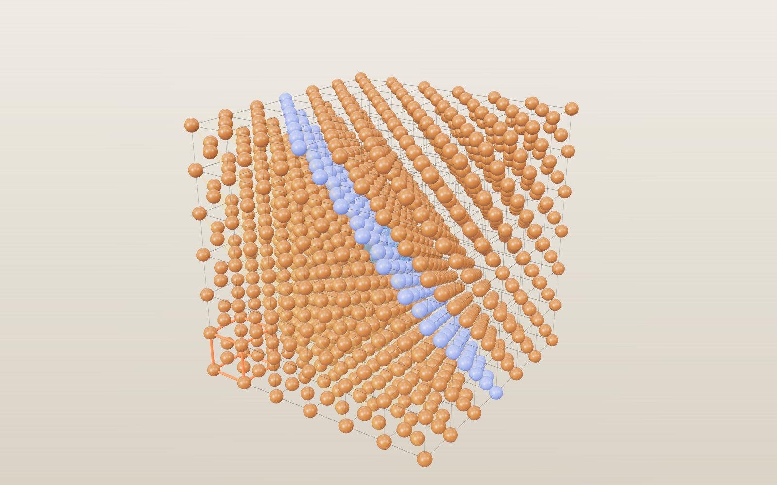
This device cannot show the 3D model. You can read the guide steps below as text.
1 / 15 One cell of saltThis cube, 0.564 nm on a side, is the unit cell of rock salt. Sodium (purple) and chlorine (green) alternate along every edge, and the labels show which is which.
Drag to turn. Scroll or pinch to zoom.
Why does salt come out of the shaker as little cubes?
Salt forms cubes because its sodium and chlorine ions repeat in a regular crystal structure with preferred planes at right angles. Glass lacks that long-range repeating pattern and breaks along curved surfaces instead. A crystal’s unit cell connects the atomic arrangement to the shape of the larger grain.
Pour some table salt onto a dark plate and look at it with a magnifying glass. Most grains are small boxes with flat faces and square corners. Nobody cut them that way. The shape comes from the way sodium and chlorine atoms pack together, and the same packing repeats so many times that it shows up at a size you can see.
This exhibit builds 41 crystals and the 14 Bravais lattices from balls and sticks, using lattice constants from mineral handbooks, semiconductor databases and the original structure papers. Each one can be seen at three sizes: a single unit cell, a block of up to 10 × 10 × 10 cells, and the outside shape the crystal grows into. On the same stage you can cut Miller planes, count the neighbours around an atom, send an X-ray beam through the planes, heat iron until its lattice changes, and put defects into a perfect crystal. The sources are listed at the end.
A crystal is one box repeated
A crystal is a solid whose atoms sit in a pattern that repeats in all three directions. To describe it, you only need one box and its contents. Copy the box, shift it by one edge length, and do the same along the other two edges, and you have the whole crystal. That box is the unit cell, and the lengths of its edges are the lattice constants.
Two things are needed to describe a crystal completely. The lattice is the grid of points where the box repeats. The basis is what sits at each point: one atom in copper, two ions in salt, a whole molecule in sugar. Change the basis and keep the lattice, and you can get a completely different material. Rock salt, periclase and galena all have the same lattice and the same kind of basis, just with different ions.
For a cubic cell one number describes the size: a = 0.564 nm for salt and a = 0.357 nm for diamond. Hexagonal cells need two numbers. The base is a rhombus with 120° and 60° corners and side a, and the height is c. For graphite a = 0.2464 nm and c = 0.6711 nm. The least symmetric cells need six numbers: three edge lengths and three angles.
The readout shows the crystal system, the Bravais lattice symbol and the space group for each crystal. The space group adds the symmetry of the basis to the symmetry of the lattice. There are 230 of them, and the model uses the operations of several (P3₂21 for quartz, R3̄c for calcite and corundum, Pa3̄ for pyrite) to place every atom from a short list of positions in the published structures.
Fourteen ways to repeat a point
In 1850 Auguste Bravais showed that there are only 14 ways to arrange points in space so that every point has exactly the same surroundings. Choose the Bravais lattice slider, or the table in the lab below the controls, to see each one. Only the lattice points are drawn: grey at the corners, red in the body centre, blue in the face centres and green in the base centres.
The 14 lattices fall into 7 crystal systems, set by the shape of the box:
- Cubic: three equal edges at right angles. Simple, body-centred and face-centred.
- Tetragonal: a square base and a different height. Simple and body-centred.
- Orthorhombic: three different edges at right angles. Simple, base-centred, body-centred and face-centred.
- Hexagonal: two equal edges at 120°, the third at right angles to both.
- Trigonal: the rhombohedral lattice, three equal edges and three equal angles that are not 90°.
- Monoclinic: one angle that is not 90°. Simple and base-centred.
- Triclinic: nothing equal and no right angles.
Why not more? Some centrings give nothing new. A face-centred tetragonal box, for example, can be redrawn as a smaller body-centred tetragonal box, so it is not counted twice. The Axis and Angle sliders let you stretch and lean the box to see where one lattice turns into another.
Why no 5-fold symmetry? Try to tile a floor with regular pentagons and gaps appear. The same holds for any lattice: only rotations by 1, 2, 3, 4 or 6 steps per turn are compatible with repeating translations. That is why the 10-fold diffraction pattern Dan Shechtman saw in 1982 was such a surprise. It came from a quasicrystal, ordered but never repeating, and it earned him the 2011 Nobel Prize in Chemistry.
From one cell to a grain to a stone
The buttons at the bottom of the stage switch between three zoom levels.
Cell shows one unit cell with balls and sticks. Atoms on the corners, edges and faces are drawn in full even though they are shared with the neighbouring cells, so a salt cell shows 27 ions but owns only 8.
Block stacks cells along each edge. For small cells such as copper or salt the block goes up to 10 × 10 × 10 cells: a thousand cells and more than 9,000 ions for salt. Every atom on screen is one copy of the same sphere, drawn in a single batch with its own position and colour, which keeps even large blocks smooth on a phone. Cells with many atoms, such as C₆₀ with 240 atoms per cell, stop at a smaller block.
Shape shows the solid the crystal grows into. Real crystals are bounded by flat faces that follow lattice planes, and the slowest-growing faces end up largest. Salt grows as cubes bounded by {100} faces. Diamond often grows as an octahedron of {111} faces. Quartz grows as a six-sided prism capped by rhombohedron faces, calcite breaks into rhombohedra of {104} faces, and pyrite forms pyritohedra with twelve five-sided faces. The model builds each shape by cutting a large box with the face planes of the crystal, each set at a distance chosen to match the common habit described in the Handbook of Mineralogy.
The readout on the Shape level shows how many unit cells fit in a grain 1 mm across. For salt it is about 5.6 × 10¹⁸, all lined up in the same orientation. That count is what turns the shape of one tiny box into the shape of a crystal you can hold.
Rock salt: two lattices pushed together
In the salt cell, sodium ions sit at the eight corners and at the centre of all six faces. That arrangement is called face-centred cubic. Chloride ions sit halfway along each of the twelve edges and one sits right in the middle. The chloride ions on their own also form a face-centred cubic lattice, shifted by half a cell edge. So a salt crystal is two face-centred cubic lattices, one of each ion, pushed into each other.
Along any edge of the cube the ions alternate: sodium, chlorine, sodium. Each sodium ion has six chloride neighbours, one in each direction along the three axes, and each chloride ion has six sodium neighbours. The distance between neighbours is half the cell edge, 0.282 nm. Shannon's table gives the ionic radii as 102 pm for Na⁺ and 181 pm for Cl⁻ in this six-fold position. Add them and you get 283 pm, so the ions touch along the edges, as the model shows when you set Ball size to 100%.
The sticks between ions in the salt model are not bonds in the sense of shared electrons. Salt is held together by the attraction between positive and negative ions. The sticks only show which ions are next to each other. Sylvite (KCl), periclase (MgO) and galena (PbS) use the same layout, which is why it is called the rock-salt structure.
How many neighbours fit
Turn on Neighbours and the model joins the nearest neighbours of one central atom into a solid, the coordination polyhedron. Four neighbours make a tetrahedron, six an octahedron, eight a cube and twelve a cuboctahedron.
For ionic crystals Linus Pauling set out a simple rule in 1929. A positive ion is usually smaller than the negative ions around it. As many negative ions crowd around it as fit without the positive ion rattling in its hole. The ratio of the radii r₊/r₋ decides the number:
| r₊/r₋ | Neighbours | Shape | Example in the model |
|---|---|---|---|
| 0.225 to 0.414 | 4 | Tetrahedron | Sphalerite ZnS (0.326) |
| 0.414 to 0.732 | 6 | Octahedron | Rock salt NaCl (0.564), MgO (0.514) |
| above 0.732 | 8 | Cube | Caesium chloride CsCl (0.961), fluorite CaF₂ (0.855) |
The radii come from Shannon's 1976 table, which gives each ion a radius for each number of neighbours. Caesium is large enough to hold 8 chloride ions, so CsCl uses a different layout from NaCl: one ion at the corners of a cube and the other in the middle. In fluorite each calcium ion sits in a cube of 8 fluoride ions, and each fluoride ion in a tetrahedron of 4 calcium ions, which is what the formula CaF₂ requires.
The rule is a guide, not a law. KCl has a radius ratio of 0.762, which points to 8 neighbours, but sylvite has 6, like salt. Corundum, Al₂O₃, has a ratio of 0.382 that points to 4, yet each aluminium ion sits in an octahedron of 6 oxygen ions. The model shows both the measured structure and the ratio, so you can see where the rule works and where it does not.
Metals: packing equal balls
A pure metal is the simplest crystal: one kind of atom, held together by electrons shared by the whole crystal. The atoms behave like equal balls, and there are only a few good ways to stack them.
Face-centred cubic (copper, aluminium, gold, γ-iron): each atom touches 12 others and the balls fill 74.05% of space, π/√18. Johannes Kepler guessed in 1611 that no packing of equal balls does better. A computer-checked proof was published in 2017.
Hexagonal close-packed (magnesium, titanium): the same layers of balls as face-centred cubic, stacked ABAB instead of ABCABC. It also fills 74.05% when c/a is 1.633. Magnesium comes close, with c/a = 1.624. Zinc is stretched along c, with c/a = 1.856, so each zinc atom has 6 close neighbours in its layer and 6 further away.
Body-centred cubic (α-iron, tungsten): an atom in the middle of a cube of 8. It fills 68.02% of space, π√3/8, slightly less than close packing.
Simple cubic fills only 52.36%, π/6. Polonium is the only element that crystallises this way at normal pressure.
Turn on Neighbours for copper and then for α-iron to compare the 12-neighbour cuboctahedron with the 8-neighbour cube. The packing fraction in the readout is calculated from the balls at contact, so it reads 74% for copper and 68% for iron.
How easily a metal bends depends on its lattice. Face-centred cubic metals have 12 easy combinations of slip plane and slip direction, so copper, aluminium and gold can be bent, drawn into wire and hammered thin. Hexagonal metals such as magnesium have far fewer at room temperature, which makes them harder to shape.
Diamond and graphite: the same atom, two ways to bond
Diamond and graphite are both pure carbon, yet one is the hardest mineral and the other is soft enough to write with. The difference is how each carbon atom uses its four outer electrons.
In diamond each carbon forms four covalent bonds, all the same length, 0.154 nm. The four neighbours sit at the corners of a tetrahedron around it. The cubic cell is a face-centred cubic lattice of carbon plus a second copy of the same lattice shifted a quarter of the way along the cube's body diagonal. Count the atoms that belong to one cell and you get eight. Because every bond is strong and they point in all directions, there is no easy direction to push the atoms apart. The balls at contact fill only 34% of space, which shows how much a covalent network is held by its bonds rather than by packing.
In graphite each carbon forms three bonds, 0.142 nm long, to neighbours in the same plane. The atoms form flat sheets of hexagons. Neighbouring sheets are 0.3355 nm apart, more than twice the bond length, and only weak forces act across that gap. The sheets are stacked in an ABAB pattern: every second sheet lines up with the first, so the cell height c is two gaps, 0.6711 nm. Switch to graphite and look at the orange marker, which shows one gap between an atom and the one directly above it.
A single sheet is graphene. In 2004 Andre Geim and Konstantin Novoselov peeled it off graphite with sticky tape, and they received the 2010 Nobel Prize in Physics. Graphene is weighed per area rather than per volume. With 2 atoms in a cell of area (√3/2)a², the model gets 0.761 mg per square metre, close to the 0.77 mg the Nobel committee quotes.
Carbon has a third common form. C₆₀, found in 1985, is a closed cage of 60 atoms shaped like a football, with 12 pentagons and 20 hexagons. In solid C₆₀ the cages stack face-centred cubic with a = 1.417 nm, about four times the cell of copper. The model draws every one of the 60 atoms in each cage. At room temperature the real molecules spin in place; below 249 K they lock into fixed orientations.
Semiconductors: silicon and its relatives
Silicon has the diamond structure with a = 0.5431 nm. It is the best-measured crystal there is: CODATA gives its lattice constant as 543.102 0511 pm. That precision made silicon the tool for one of the most careful measurements in physics. The Avogadro constant was found by counting the atoms in a polished sphere of silicon-28: measure the sphere's mass, its volume and the cell size, and the density formula run backwards gives the number of atoms in a mole.
Germanium has the same structure with a = 0.5658 nm. The first transistor, built in 1947, used a germanium crystal. Replace one kind of atom on every second site and you get the zinc-blende structure of gallium arsenide (GaAs) and silicon carbide (SiC), which are used in lasers, fast radio chips and power electronics. Gallium nitride crystallises in the hexagonal wurtzite structure, still with every atom in a tetrahedron of the other kind. Good GaN crystals made bright blue LEDs possible, recognised by the 2014 Nobel Prize in Physics.
A semiconductor is useful because a trace of the right impurity changes how it conducts. Choose the Substitution defect for silicon and three silicon atoms become phosphorus. Phosphorus has one more outer electron than silicon, and that electron is free to carry current. Boron, with one fewer, leaves a hole. Chip silicon is first purified to better than 99.9999999% and then doped on purpose.
Silicon for chips is grown as a single crystal by the method Jan Czochralski found in 1916: a seed crystal touches molten silicon and is pulled up slowly while turning. The result is a round ingot, which is why the Shape level shows silicon as a cylinder rather than a polyhedron. The ingot is sawn into wafers.
Minerals built from polyhedra
Many minerals are easier to understand as linked polyhedra than as balls.
Quartz, SiO₂, is built from SiO₄ tetrahedra, each silicon 0.161 nm from four oxygens. Every oxygen is shared by two tetrahedra, so the network is joined at every corner. The tetrahedra form spirals along the c axis, and the spirals turn either left or right, giving left- and right-handed quartz. The model uses the positions refined by Levien, Prewitt and Weidner in 1980.
Calcite, CaCO₃, has flat carbonate groups, a carbon with three oxygens 0.128 nm away, in layers between calcium ions. Each calcium has 6 oxygen neighbours. The layered, lopsided arrangement slows light differently in different directions, which gives calcite its strong double refraction.
Corundum, Al₂O₃, is a close-packed stack of oxide ions with aluminium in two thirds of the octahedral gaps. Pure corundum is colourless. A little chromium in place of aluminium makes it red, and the stone is called ruby; iron and titanium make it blue, and any gem corundum that is not red is called sapphire. Choose Substitution for corundum to put chromium into the lattice.
Rutile, TiO₂, has chains of TiO₆ octahedra running along c. Most titanium dioxide ends up as the white pigment in paint, paper and plastics.
Pyrite, FeS₂, looks like rock salt with each chloride ion replaced by a pair of sulfur atoms 0.216 nm apart. The pairs point along the four body diagonals, which lowers the symmetry and explains the striped cubes and twelve-faced pyritohedra of fool's gold.
Perovskites, with the formula ABX₃, put a small ion (titanium in SrTiO₃) at the centre of an octahedron of oxygens and a large ion (strontium) in the big gap between octahedra, with 12 oxygen neighbours. Strontium titanate is the ideal cubic version. Solar cells made from perovskite crystals rose from about 4% efficiency in 2009 to the level of silicon cells in research labs.
Lithium cobalt oxide, LiCoO₂, stacks sheets of CoO₂ with lithium ions between them. When a lithium-ion battery charges, lithium leaves these gaps; when it discharges, it returns. John Goodenough's group described this material as a cathode in 1980.
Ice: a crystal held by hydrogen bonds
Ordinary ice is called ice Ih, with h for hexagonal. Each water molecule's oxygen has four oxygen neighbours at about 0.276 nm, arranged at the corners of a tetrahedron, much like the carbon atoms in diamond. Between each pair of neighbouring oxygens lies exactly one hydrogen. It is covalently bonded to one oxygen, 99 pm away, and held to the other by a hydrogen bond, 177 pm away.
Each molecule gives two hydrogens to its neighbours and receives two. The model places the hydrogens so that this rule holds for every molecule in the block. In real ice which hydrogen sits near which oxygen is disordered and changes over time; the model shows one allowed arrangement.
The tetrahedral pattern leaves an open structure of puckered hexagonal rings with empty channels running through them. When ice melts, part of that network collapses and the molecules pack more closely. That is why ice, at 0.9167 g/cm³ at 0 °C, floats on water, which is 0.99984 g/cm³ at the same temperature (NIST).
Ice has more than one crystal form. Ice Ic keeps the same four hydrogen bonds per molecule but stacks the layers like diamond instead of like a hexagonal crystal, with a cubic cell of 0.6358 nm. It forms when tiny droplets freeze at the very low temperatures of the high atmosphere. Ice VII is two cubic ice networks passing through each other without touching. It needs a pressure above about 2 GPa. Its density is about 1.65 g/cm³, nearly twice that of ordinary ice, and in 2018 it was found trapped inside diamonds from the deep mantle.
Molecules as building blocks
In sugar and proteins the basis is a whole molecule, and the bonds inside the molecule are much stronger than the forces between molecules. The model draws each molecule as one ball to keep the lattice visible.
A sucrose crystal is monoclinic, with 2 molecules per cell. One molecule is related to the other by a twofold screw axis, a half turn plus a shift of half a cell. The cell is 1.086 × 0.871 × 0.776 nm with β = 102.9°, and the density formula gives 1.590 g/cm³ against 1.587 measured.
Protein crystals are much larger. The tetragonal crystals of lysozyme, an enzyme from egg white, have a = 7.91 nm and c = 3.79 nm, with 8 molecules per cell. If the cell held only protein its density would be 0.80 g/cm³, less than water. The rest is water: Brian Matthews found in 1968 that protein crystals have about 2.2 ų of cell volume per dalton of protein, which leaves close to half the crystal as solvent. Lysozyme was the first enzyme whose structure was solved, in 1965, and protein crystallography is still how most enzyme structures are found.
Naming planes: Miller indices
A plane through a crystal is named by three whole numbers (h k l), its Miller indices. Take the plane closest to the origin that cuts the cell edges at a/h, b/k and c/l. A zero means the plane runs parallel to that edge. So (1 0 0) is a cube face, (1 1 0) cuts two edges and runs along the third, and (1 1 1) cuts off a corner. Braces such as {100} mean the whole family of planes equivalent by symmetry, and in a cube {100} has three members.
Turn on Miller plane and set h, k and l. The model draws the parallel planes h x + k y + l z = whole number through the block and colours the atoms that sit on the middle one. The readout gives the spacing between planes, d, and the number of atoms per square nanometre on one plane.
For a cubic crystal the spacing is d = a / √(h² + k² + l²). For copper, (1 1 1) planes are 0.3615 / √3 = 0.2087 nm apart, and each holds 17.67 atoms per square nanometre in a pattern of triangles. That is the densest plane in a face-centred cubic metal, which is why metals slip along {111}. In body-centred iron the densest planes are {110} instead. For other crystal systems the model computes d from the reciprocal lattice, which works for any cell shape.
Where crystals split
When you hit a crystal, it tends to break along planes where the fewest or weakest bonds cross. Mineralogists call this cleavage. The Handbook of Mineralogy lists perfect cleavage on {001} for halite, the mineral name for rock salt. In a cube there are three such planes, at right angles to each other. Split salt along all three and each piece is again a box with flat faces and square corners. This is why salt grains look like cubes.
Diamond has perfect cleavage on {111}, planes that cut across the cube's corners at an angle. Turn on Split with diamond and watch the bonds: where the block comes apart, only one bond per atom crosses the plane, while atoms on either side are joined by three. Gem cutters use this to split rough stones before sawing and polishing.
Graphite cleaves between its sheets. Only weak forces cross there, so the sheets slide and flake off easily, which is how a pencil leaves a mark on paper. Fluorite cleaves on {111}, so a fluorite cube can be split down to an octahedron. Calcite cleaves on {104} in three directions that are not at right angles, which is why every broken piece is a rhombohedron. Quartz has no good cleavage at all: its tetrahedra are linked in every direction, and it breaks along curved, shell-like surfaces.
The model chooses the split among the planes of the cleavage family that cross the middle of the block, using the widest gap between atomic layers. For diamond that is the gap between double layers, crossed by one bond per atom.
X-ray diffraction and Bragg's law
The spacing of atoms in a crystal is close to the wavelength of X-rays, so a crystal scatters X-rays in a pattern that depends on its structure. In 1913 William Lawrence Bragg explained it with a picture of reflection. X-rays reflect weakly from each plane of atoms. The reflections from neighbouring planes add up only when the extra path of the deeper ray, 2 d sin θ, is a whole number of wavelengths:
n λ = 2 d sin θ
Here θ is the angle between the beam and the planes, d is the plane spacing and n is a whole number, the order. The reflected beam leaves at 2θ from the incoming one, so diffractometers report 2θ.
Turn on X-ray beam. The purple beam comes from the tube on the left, meets the chosen (h k l) planes and leaves towards the detector. Move the Angle 2θ slider. The detector stays dark until the angle meets Bragg's law, then the yellow beam lights. For salt and copper Kα X-rays (λ = 1.5406 Å), the (2 0 0) planes are 0.2820 nm apart and reflect at 2θ = 31.70°. Change the wavelength to molybdenum Kα, 0.7093 Å, and the same planes reflect at a smaller angle, 14.45°.
The small graph in the readout is the powder pattern: what a diffractometer records from a powder in which tiny crystals point in every direction, so every set of planes reflects at once. Each line is one family of planes. The model computes the positions exactly from the cell and Bragg's law. The heights come from the structure factor, the sum of the waves scattered by every atom in the cell with their phases, times the number of equivalent planes and an angle factor. For salt with copper Kα the strongest lines are (2 0 0) at 31.70°, (2 2 0) at 45.45° and (2 2 2) at 56.47°, as in the measured pattern.
Some planes never reflect, however you turn the crystal. In a face-centred cubic lattice the atoms in the face centres send waves that exactly cancel the corner atoms for (1 0 0), so the first copper line is (1 1 1) at 43.32°. In diamond and silicon the second copy of the lattice also cancels (2 0 0). These missing reflections, called systematic absences, are how crystallographers tell the lattice type from a pattern. Try (1 0 0) on copper: the readout says the reflection is absent and the beam never lights.
Potassium chloride shows a subtler case. K⁺ and Cl⁻ both have 18 electrons, so to X-rays they look almost the same. The (1 1 1) reflection, which depends on the difference between the two ions, nearly vanishes, and the pattern looks like that of a simple cubic crystal with half the cell. In salt, Na⁺ has 10 electrons and Cl⁻ 18, so (1 1 1) shows clearly.
Iron at 912 °C and other phase changes
The Temperature slider runs from −200 to 1600 °C. At higher temperature the atoms shake harder around their places, and the model shows that as a jiggle that grows with temperature. Past the melting point the crystal loses its order: atoms still touch but no longer sit on a lattice.
Iron also changes its lattice on the way up. Below 912 °C it is body-centred cubic, α-iron or ferrite. Between 912 and 1394 °C it is face-centred cubic, γ-iron or austenite. From 1394 °C to melting at 1538 °C it is body-centred cubic again, δ-iron. The face-centred form packs the atoms more closely, so iron shrinks slightly when it changes at 912 °C. Its larger gaps can also hold much more carbon. Steel is heat treated by using this: heat it into the γ range, where carbon dissolves, then cool it at the right speed to trap the carbon in different arrangements.
Tin changes the other way. Above 13.2 °C the stable form is white tin, a tetragonal metal. Below that, grey tin with the diamond structure is stable. The change is slow and needs a long cold spell, but when it happens the tin swells by about a quarter and crumbles. Old stories blame this tin pest for lost buttons and organ pipes; most are hard to confirm.
Ice melts at 0 °C. The model switches crystals with a temperature family automatically, so moving the slider on iron or tin shows each phase in turn. The lattice constants for γ- and δ-iron are measurements made at high temperature by Basinski, Hume-Rothery and Sutton in 1955.
Defects: why real crystals are never perfect
No real crystal is perfect, and many useful properties come from the faults. The Defect slider works on the Block level.
- Vacancy: an atom missing from its site. In salt a sodium and a chloride ion go missing together, so the crystal stays neutral. Vacancies let atoms move through a solid, which is how diffusion works.
- Interstitial: an extra atom squeezed into a gap between sites. In iron the model puts a carbon atom into the largest gap. That is the start of steel.
- Substitution: a foreign atom on a normal site. Zinc in copper makes brass, chromium in corundum makes ruby, boron in diamond makes the Hope Diamond blue, and phosphorus in silicon makes a transistor work.
- Edge dislocation: an extra half plane of atoms that ends inside the crystal. Look along the red ⊥ marker and you will see the planes above bend around the end of the extra one. A pure metal yields at a stress thousands of times lower than a perfect crystal would need, because a dislocation lets the planes slip one row at a time. G. I. Taylor, Egon Orowan and Michael Polanyi proposed this independently in 1934.
- Twin boundary: one part of the crystal is the mirror image of the other across a plane. In the model the half above a (1 1 1) plane is reflected, which in a face-centred cubic metal gives the coherent twin seen in brass and copper.
The dislocation in the model is placed with the displacement field of elasticity theory, the standard way to build one in an atomic model, with a Burgers vector of one cell edge.
Checking the densities
If you know what is in one cell and how big the cell is, you know the density. The mass of one cell is Z · M / N_A, where Z is the number of formula units the cell owns, M is the molar mass and N_A is the Avogadro constant, 6.022 140 76 × 10²³ per mole (exact by definition). Divide by the cell volume and you have ρ = Z · M / (N_A · V). For a cubic cell V = a³; for a hexagonal cell V = (√3 / 2) · a² · c; in general V is the volume of the box made by the three edge vectors.
For salt, Z = 4 and M = 22.990 + 35.45 = 58.44 g/mol (CIAAW atomic weights). The cell volume is (0.56404 nm)³ = 0.17944 nm³, or 1.7944 × 10⁻²² cm³. That gives ρ = 4 × 58.44 / (6.022 × 10²³ × 1.7944 × 10⁻²²) = 2.163 g/cm³. The Handbook gives a measured density of 2.168, so the two differ by 0.2%. The lab under the controls repeats this sum for whichever crystal is on the stage.
| Crystal | Cell | Z | Calculated ρ (g/cm³) | Measured (g/cm³) |
|---|---|---|---|---|
| Rock salt NaCl | a = 0.56404 nm | 4 | 2.163 | 2.168 |
| Caesium chloride CsCl | a = 0.4123 nm | 1 | 3.989 | 3.988 |
| Fluorite CaF₂ | a = 0.54626 nm | 4 | 3.181 | 3.175 to 3.184 |
| Diamond C | a = 0.35668 nm | 8 | 3.516 | 3.511 |
| Graphite C | a = 0.2464, c = 0.6711 nm | 4 | 2.261 | 2.09 to 2.23 |
| Silicon Si | a = 0.54310 nm | 8 | 2.329 | 2.329 |
| Copper Cu | a = 0.36149 nm | 4 | 8.935 | 8.95 |
| α-iron Fe | a = 0.28665 nm | 2 | 7.874 | 7.874 |
| Tungsten W | a = 0.31652 nm | 2 | 19.254 | 19.25 |
| Magnesium Mg | a = 0.32094, c = 0.52108 nm | 2 | 1.737 | 1.738 |
| Quartz SiO₂ | a = 0.49135, c = 0.5405 nm | 3 | 2.649 | 2.65 |
| Calcite CaCO₃ | a = 0.49896, c = 1.7061 nm | 6 | 2.711 | 2.710 |
| Pyrite FeS₂ | a = 0.54179 nm | 4 | 5.010 | 5.018 |
| Ice Ih H₂O | a = 0.4518, c = 0.7356 nm | 4 | 0.920 | 0.9196 at −20 °C |
| Sucrose | 1.0863 × 0.8705 × 0.7759 nm, β 102.9° | 2 | 1.590 | 1.587 |
Graphite's measured range is lower than the calculated value because natural graphite contains pores and stacking faults; the Handbook's own calculated value is 2.26. Ice shrinks slightly as it cools, so its density depends on temperature. NIST lists 0.9167 g/cm³ at 0 °C and 0.9196 at −20 °C.
The readout also shows a second number, ρ drawn. That is what you get if you add up the mass of every ball on screen and divide by the block volume. For one salt cell it gives 7.24 g/cm³, more than three times too much. The reason is sharing. An ion on a corner belongs to eight cells, one on an edge to four and one on a face to two, so only a fraction of it counts for this cell. One salt cell draws 27 ions but owns only 8 of them: 8 × ⅛ + 6 × ½ = 4 sodium ions and 12 × ¼ + 1 = 4 chloride ions. As you stack more cells, a smaller share of the ions sits on the outside of the block, and ρ drawn falls towards the true value: 4.22 at 2 × 2 × 2, 3.08 at 4 × 4 × 4 and 2.50 at 10 × 10 × 10. A grain of salt half a millimetre wide is about 890,000 cells across, so in real salt the surface share is tiny.
What the model leaves out
The balls are drawn smaller than the atoms so you can see the structure; Ball size at 100% uses Shannon's ionic radii for ionic crystals and half the nearest-neighbour distance for the others. Molecules in sugar and lysozyme are single balls. Real atoms vibrate around their positions, by more at higher temperature, which is also why the lattice constants change a little with temperature; the model keeps room-temperature cells except for the high-temperature forms of iron. The X-ray intensities use one smooth fall-off with angle for every atom, so the heights of the lines are close but not exact, while their positions are exact. The C₆₀ molecules are frozen in one orientation. Shapes on the Shape level are typical habits, and real crystals grow many variations. Ice in the model has one fixed arrangement of hydrogens instead of a constantly changing one.
How crystal structures were first measured
Nicolas Steno noticed in 1669 that quartz crystals of every size meet at the same angles between matching faces. In the same year Erasmus Bartholin saw a double image through Iceland spar, a clear calcite. René-Just Haüy found that broken calcite always came apart into smaller rhombohedra and proposed in his 1784 book that crystals are stacks of identical small blocks. Bravais counted the possible lattices in 1850, and by the end of the century the 230 space groups were known.
Nobody could see the atoms until 1912, when Max von Laue's group sent X-rays through a crystal and recorded a pattern of spots. It showed at once that X-rays are waves and that crystals are regular grids. William Henry Bragg and his son William Lawrence Bragg turned the spots into structures: salt in 1913, then diamond. They shared the 1915 Nobel Prize in Physics. Over the following decades the same method solved minerals, metals and then the molecules of life: Dorothy Hodgkin's penicillin and vitamin B₁₂, the X-ray patterns of DNA fibres recorded by Rosalind Franklin and Raymond Gosling in 1952, and the first enzyme, lysozyme, in 1965. The lattice constants used here come from the same kind of measurement, refined over the following century.
Questions
Why does salt break into cubes but glass does not?
Salt is a crystal, so its weakest planes run straight through the whole grain and all three are at right angles. Glass has no repeating pattern, so there are no preferred planes and it breaks along curved surfaces.
What does Z mean in the density formula?
Z is the number of formula units that one unit cell owns after sharing is counted. For salt it is 4 NaCl units, for diamond 8 carbon atoms, for graphite 4 carbon atoms and for ice 4 water molecules.
Why is the ρ drawn number so high for small blocks?
It counts every ball you see, including the ones on corners, edges and faces that also belong to the next cell. A small block is mostly surface, so the overcount is large. It shrinks as the block grows.
Diamond and graphite are both carbon. Why is graphite less dense?
Graphite's sheets are 0.3355 nm apart, which leaves a lot of empty space between them. In diamond every atom is bonded to four neighbours in all directions, so the atoms pack more tightly: 3.52 g/cm³ against 2.26.
Why does ice float?
Each water molecule in ice is hydrogen bonded to exactly four neighbours, which forces an open arrangement of hexagonal rings. Liquid water packs the molecules more closely, so it is denser: 0.99984 g/cm³ at 0 °C against 0.9167 for ice.
Are the colours of the atoms real?
No. Atoms have no colour of their own at this size. The model uses common teaching colours, such as purple sodium, green chlorine, red oxygen and white hydrogen, and copper and gold balls in their metal colours.
How big is one salt grain in unit cells?
A grain 0.5 mm wide is 500,000 nm across. Divided by 0.564 nm, that is about 890,000 cells along each edge, or around 7 × 10¹⁷ cells in the whole grain.
Why are there only 14 Bravais lattices?
A lattice must look the same from every point. Once the 7 box shapes are fixed, only some ways of adding extra points keep that true, and several of the others turn out to be the same lattice drawn with a different box. Bravais showed in 1850 that 14 remain.
What is the difference between a lattice and a crystal structure?
The lattice is the grid of repeat points. The crystal structure is the lattice plus the atoms placed at each point. Copper and γ-iron share the face-centred cubic lattice with one atom per point; salt uses the same lattice with a pair of ions per point.
Why does the detector stay dark for some planes even at the right angle?
The waves from different atoms in the cell can cancel. In a face-centred cubic lattice that happens for every (h k l) that mixes odd and even numbers, so (1 0 0) and (1 1 0) never reflect from copper. The readout says so when you pick such a plane.
Why does iron change structure when it is heated?
Each structure has a slightly different energy and a slightly different amount of atomic jiggling. At 912 °C the face-centred form becomes the more stable one, and the iron rearranges.
Is glass a crystal?
No. Glass is amorphous: its atoms have the same kind of neighbours as in quartz, but the pattern does not repeat over long distances. That is why it has no cleavage, no flat crystal faces and no sharp X-ray lines.
How do you see a dislocation in a real metal?
With a transmission electron microscope. The strain around a dislocation bends the electron beam, and dislocations show up as dark lines in thin foils. Their number per area rises sharply when a metal is bent or hammered, which is why it gets harder.
Can a crystal have fivefold symmetry?
Not a periodic one. Pentagons cannot fill a plane without gaps, and the same argument rules out fivefold axes in any lattice. Quasicrystals show 5-fold and 10-fold diffraction patterns because they are ordered without repeating.
Terms
- Unit cell: the smallest box whose copies, stacked along its edges, rebuild the whole crystal.
- Lattice constant: the length of a unit cell edge, a for cubic cells, a and c for hexagonal ones.
- Lattice and basis: the grid of repeat points, and the atoms placed at each point.
- Bravais lattice: one of the 14 ways to repeat a point in space so every point has the same surroundings.
- Crystal system: the shape class of the cell; there are 7.
- Space group: the full symmetry of a crystal structure; there are 230.
- Face-centred cubic: a cubic lattice with a point at each corner and in the middle of each face.
- Body-centred cubic: a cubic lattice with a point at each corner and one in the middle.
- Coordination number: how many nearest neighbours an atom has.
- Packing fraction: the share of space filled by atoms drawn as touching balls.
- Miller indices (h k l): three whole numbers that name a family of parallel planes.
- Cleavage: the tendency of a crystal to split along flat planes where its bonds are weakest.
- Bragg's law: n λ = 2 d sin θ, the condition for X-rays reflected by parallel planes to add up.
- Systematic absence: a reflection that is always missing because the waves from the cell cancel.
- Formula unit (Z): the number of NaCl units, atoms or molecules that one cell owns.
- Dislocation: a line defect where an extra half plane of atoms ends inside the crystal.
- Habit: the outside shape a crystal usually grows into.
Sources
- Mineralogical Society of America, Handbook of Mineralogy: Halite, Sylvite, Periclase, Galena, Fluorite, Sphalerite, Diamond, Graphite, Quartz, Calcite, Corundum, Rutile, Pyrite, Gold, Copper (cell data, densities, cleavage, habit). https://www.handbookofmineralogy.org/ (checked 18 October 2026)
- WebElements (M. Winter, University of Sheffield), crystal structures of copper, aluminium, gold, iron, tungsten, polonium, magnesium, zinc, titanium and tin. https://www.webelements.com/copper/crystal_structure.html (checked 18 October 2026)
- Ioffe Institute, NSM semiconductor archive: basic parameters of Si, Ge, GaAs, SiC and GaN. http://www.ioffe.ru/SVA/NSM/Semicond/ (checked 18 October 2026)
- NIST, CODATA value: lattice parameter of silicon. https://physics.nist.gov/cgi-bin/cuu/Value?asil (checked 18 October 2026)
- NIST, CODATA value: Avogadro constant. https://physics.nist.gov/cgi-bin/cuu/Value?na (checked 18 October 2026)
- IUPAC Commission on Isotopic Abundances and Atomic Weights (CIAAW), Standard atomic weights. https://www.ciaaw.org/atomic-weights.htm (checked 18 October 2026)
- R. D. Shannon (1976), Revised effective ionic radii and systematic studies of interatomic distances in halides and chalcogenides, Acta Crystallographica A 32, 751. https://doi.org/10.1107/S0567739476001551 (checked 18 October 2026)
- L. Pauling (1929), The principles determining the structure of complex ionic crystals, Journal of the American Chemical Society 51, 1010. https://doi.org/10.1021/ja01379a006 (checked 18 October 2026)
- L. Levien, C. T. Prewitt, D. J. Weidner (1980), Structure and elastic properties of quartz at pressure, American Mineralogist 65, 920. http://www.minsocam.org/ammin/AM65/AM65_920.pdf (checked 18 October 2026)
- American Mineralogist Crystal Structure Database (atomic positions of calcite, corundum, rutile, pyrite). https://rruff.geo.arizona.edu/AMS/amcsd.php (checked 18 October 2026)
- Z. S. Basinski, W. Hume-Rothery, A. L. Sutton (1955), The lattice expansion of iron, Proceedings of the Royal Society A 229, 459. https://doi.org/10.1098/rspa.1955.0102 (checked 18 October 2026)
- University of Cambridge, DoITPoMS teaching and learning packages: Crystallography, Miller indices, X-ray diffraction techniques, Introduction to dislocations, Phase diagrams, Slip in single crystals. https://www.doitpoms.ac.uk/tlplib/ (checked 18 October 2026)
- W. L. Bragg and W. H. Bragg (1913), The reflection of X-rays by crystals, Proceedings of the Royal Society A 88, 428. https://doi.org/10.1098/rspa.1913.0040 (checked 18 October 2026)
- W. H. Bragg and W. L. Bragg (1913), The structure of the diamond, Proceedings of the Royal Society A 89, 277. https://doi.org/10.1098/rspa.1913.0084 (checked 18 October 2026)
- NobelPrize.org: Physics 1914 (Laue), Physics 1915 (the Braggs), Chemistry 1964 (Hodgkin), Chemistry 1996 (fullerenes), Physics 2010 (graphene), Chemistry 2011 (quasicrystals), Physics 2014 (blue LEDs), Chemistry 2019 (lithium-ion batteries). https://www.nobelprize.org/ (checked 18 October 2026)
- G. I. Taylor (1934), The mechanism of plastic deformation of crystals, Proceedings of the Royal Society A 145, 362. https://doi.org/10.1098/rspa.1934.0106 (checked 18 October 2026)
- R. E. Franklin and R. G. Gosling (1953), Molecular configuration in sodium thymonucleate, Nature 171, 740. https://doi.org/10.1038/171740a0 (checked 18 October 2026)
- C. C. F. Blake et al. (1965), Structure of hen egg-white lysozyme, Nature 206, 757. https://doi.org/10.1038/206757a0 (checked 18 October 2026)
- B. W. Matthews (1968), Solvent content of protein crystals, Journal of Molecular Biology 33, 491. https://doi.org/10.1016/0022-2836(68)90205-2 (checked 18 October 2026)
- G. M. Brown and H. A. Levy (1973), Further refinement of the structure of sucrose based on neutron-diffraction data, Acta Crystallographica B 29, 790. https://doi.org/10.1107/S0567740873003353 (checked 18 October 2026)
- D. Shechtman et al. (1984), Metallic phase with long-range orientational order and no translational symmetry, Physical Review Letters 53, 1951. https://doi.org/10.1103/PhysRevLett.53.1951 (checked 18 October 2026)
- P. A. Heiney et al. (1991), Orientational ordering transition in solid C60, Physical Review Letters 66, 2911. https://doi.org/10.1103/PhysRevLett.66.2911 (checked 18 October 2026)
- T. Hales et al. (2017), A formal proof of the Kepler conjecture, Forum of Mathematics, Pi 5. https://doi.org/10.1017/fmp.2017.1 (checked 18 October 2026)
- Y. Azuma et al. (2015), Improved measurement results for the Avogadro constant using a 28Si-enriched crystal, Metrologia 52, 360. https://doi.org/10.1088/0026-1394/52/2/360 (checked 18 October 2026)
- R. Uecker (2014), The historical development of the Czochralski method, Journal of Crystal Growth 401, 7. https://doi.org/10.1016/j.jcrysgro.2013.11.095 (checked 18 October 2026)
- K. Mizushima, P. C. Jones, P. J. Wiseman, J. B. Goodenough (1980), LixCoO2: a new cathode material for batteries of high energy density, Materials Research Bulletin 15, 783. https://doi.org/10.1016/0025-5408(80)90012-4 (checked 18 October 2026)
- NIST, A. H. Harvey, Properties of Ice and Supercooled Water (densities of ice Ih and water). https://tsapps.nist.gov/publication/get_pdf.cfm?pub_id=926353 (checked 18 October 2026)
- University College London, G. A. Tribello, Structure and energy relationships in ice and crystalline hydrates, PhD thesis, 2007 (ice Ih a = 4.518 Å, c = 7.356 Å). https://discovery.ucl.ac.uk/1444691/1/U592000.pdf (checked 18 October 2026)
- London South Bank University, M. Chaplin, Water structure and science: cubic ice and ice-seven. https://water.lsbu.ac.uk/water/cubic_ice.html (checked 18 October 2026)
- B. J. Murray, D. A. Knopf, A. K. Bertram (2005), The formation of cubic ice under conditions relevant to Earth's atmosphere, Nature 434, 202. https://doi.org/10.1038/nature03403 (checked 18 October 2026)
- O. Tschauner et al. (2018), Ice-VII inclusions in diamonds: evidence for aqueous fluid in Earth's deep mantle, Science 359, 1136. https://doi.org/10.1126/science.aao3030 (checked 18 October 2026)
- IUCr Newsletter 30(4), J.-C. Boulliard, D. Cabaret, P. Giura, René-Just Haüy and the birth of crystallography. https://www.iucr.org/news/newsletter/volume-30/number-4/rene-just-hauy-and-the-birth-of-crystallography (checked 18 October 2026)
- GIA, Ruby and Sapphire descriptions. https://www.gia.edu/ruby (checked 18 October 2026)
- USGS, Mineral Commodity Summaries 2024: Salt, Potash, Silicon, Titanium mineral concentrates, Fluorspar. https://pubs.usgs.gov/periodicals/mcs2024/mcs2024-salt.pdf (checked 18 October 2026)
More exhibits
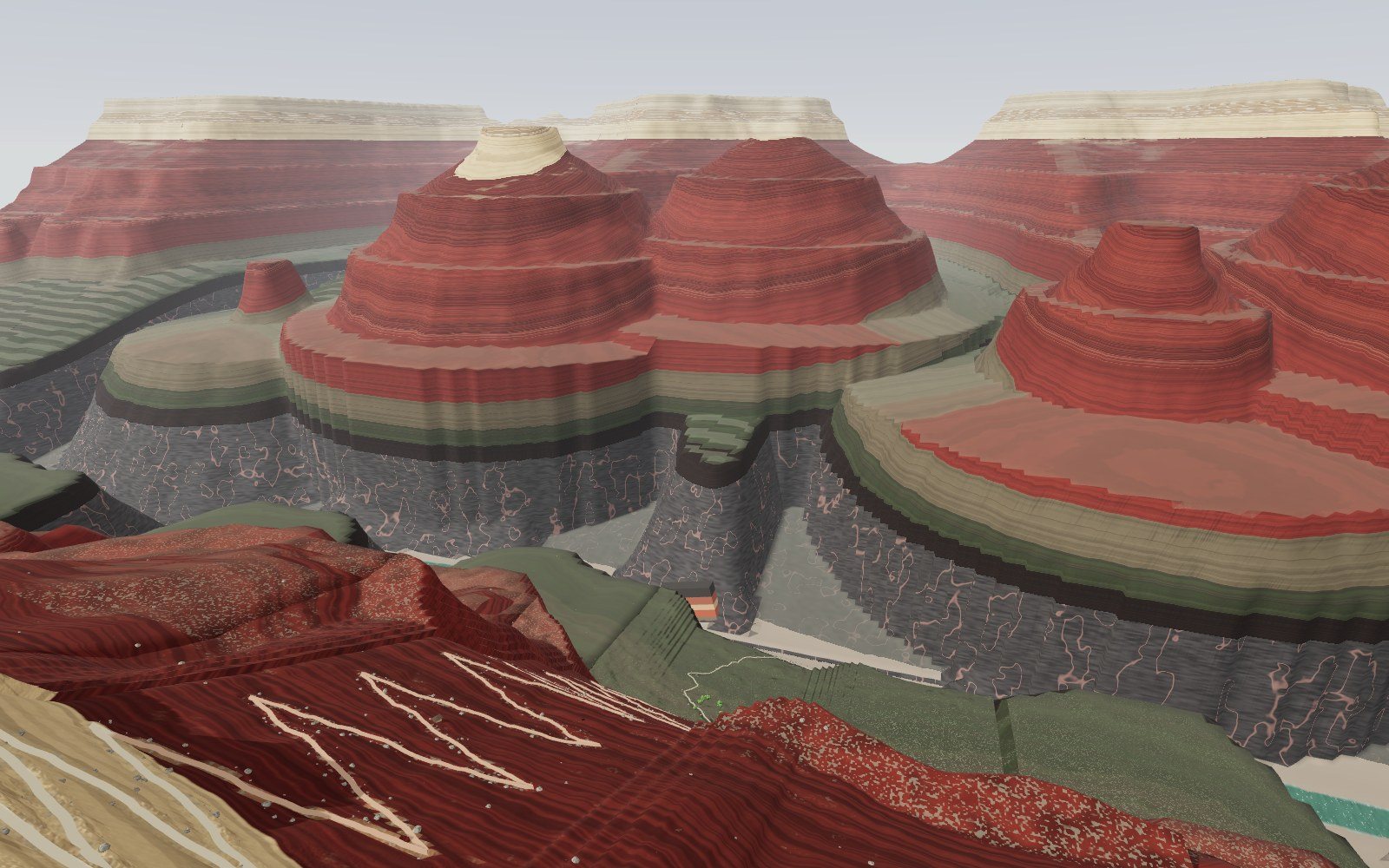
Walk down the Grand Canyon rock layers
Walk 1,360 m down the Bright Angel Trail through 13 rock layers, from a 270-million-year-old sea floor on the rim to 1.75-billion-year-old schist by the river.
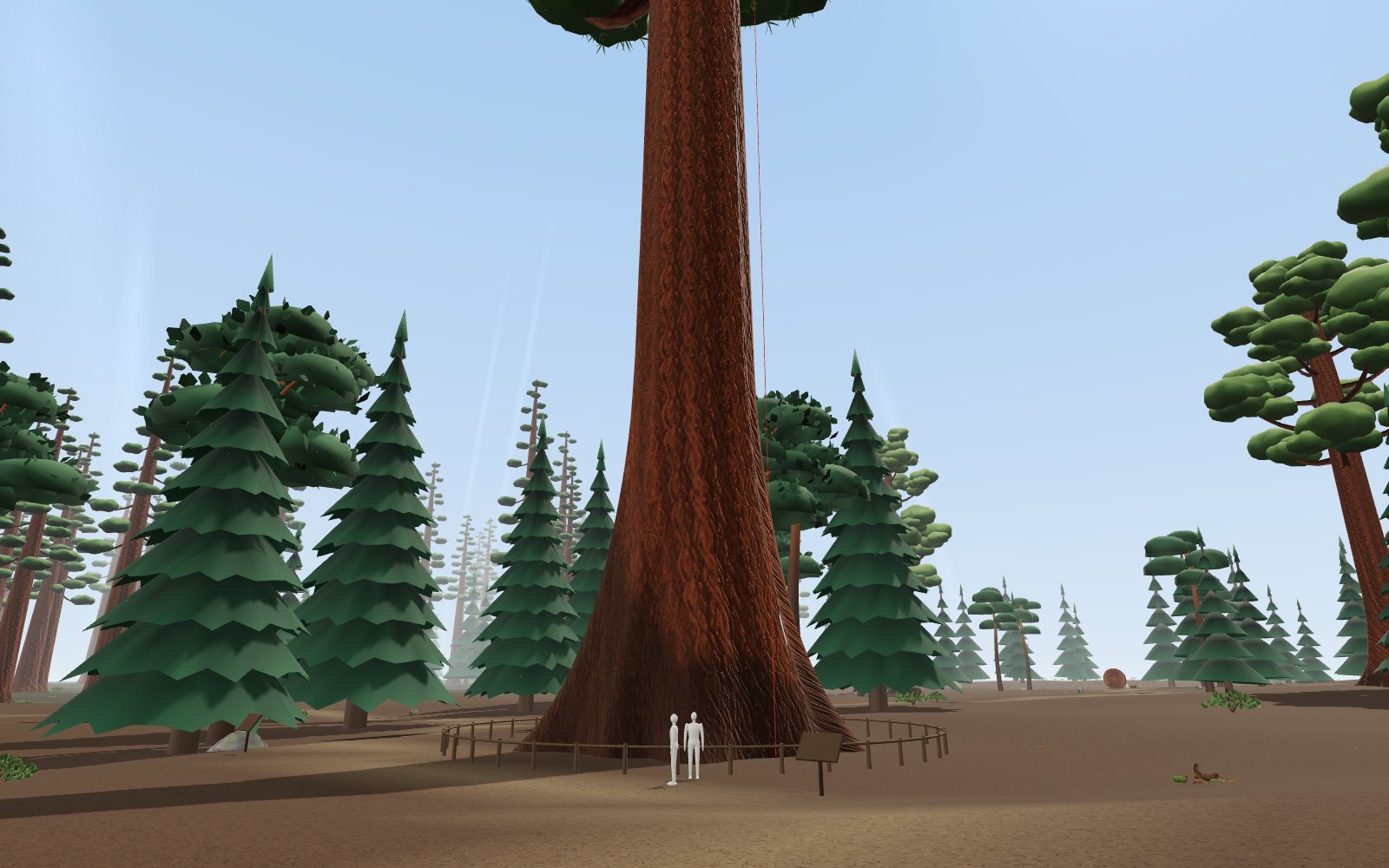
Giant sequoia forest: walk under the largest trees
Stand at the foot of the General Sherman Tree, 83.8 m tall and about 2,200 years old, then climb its trunk and see how hard the water up there is pulled.
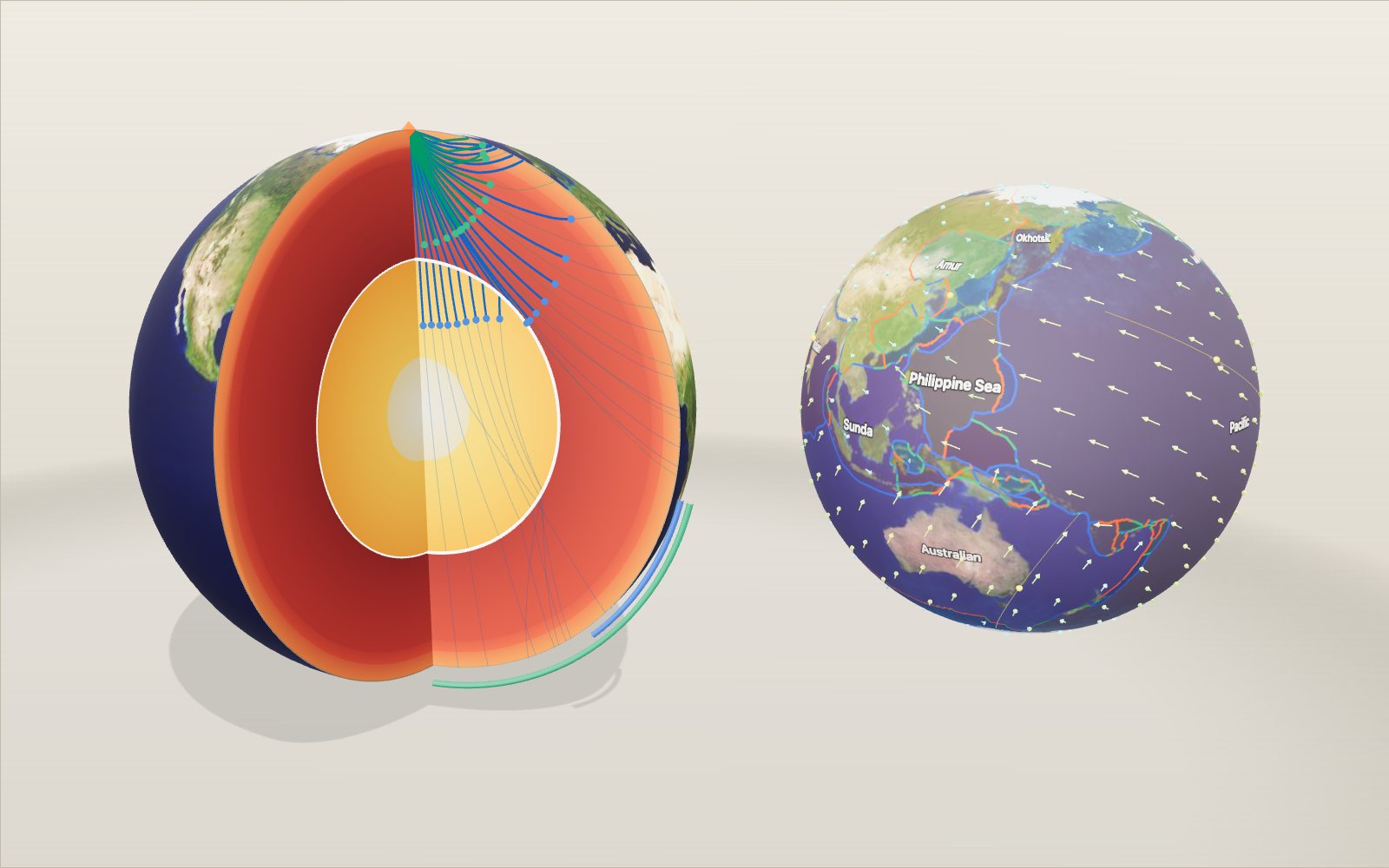
Layers of the Earth: depth and density, interactive model
Nobody has drilled deeper than 12.3 km, yet we know the pressure at the centre to within a few per cent, because earthquakes shine through the planet.
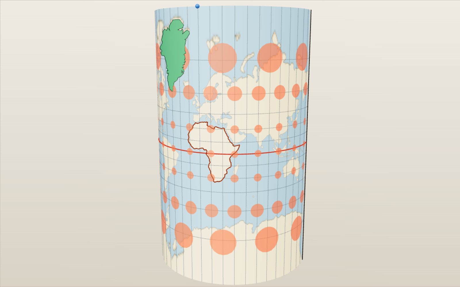
Map projections: why Greenland looks as big as Africa
On a Mercator map Greenland looks bigger than Africa, which is in fact about 14 times larger.
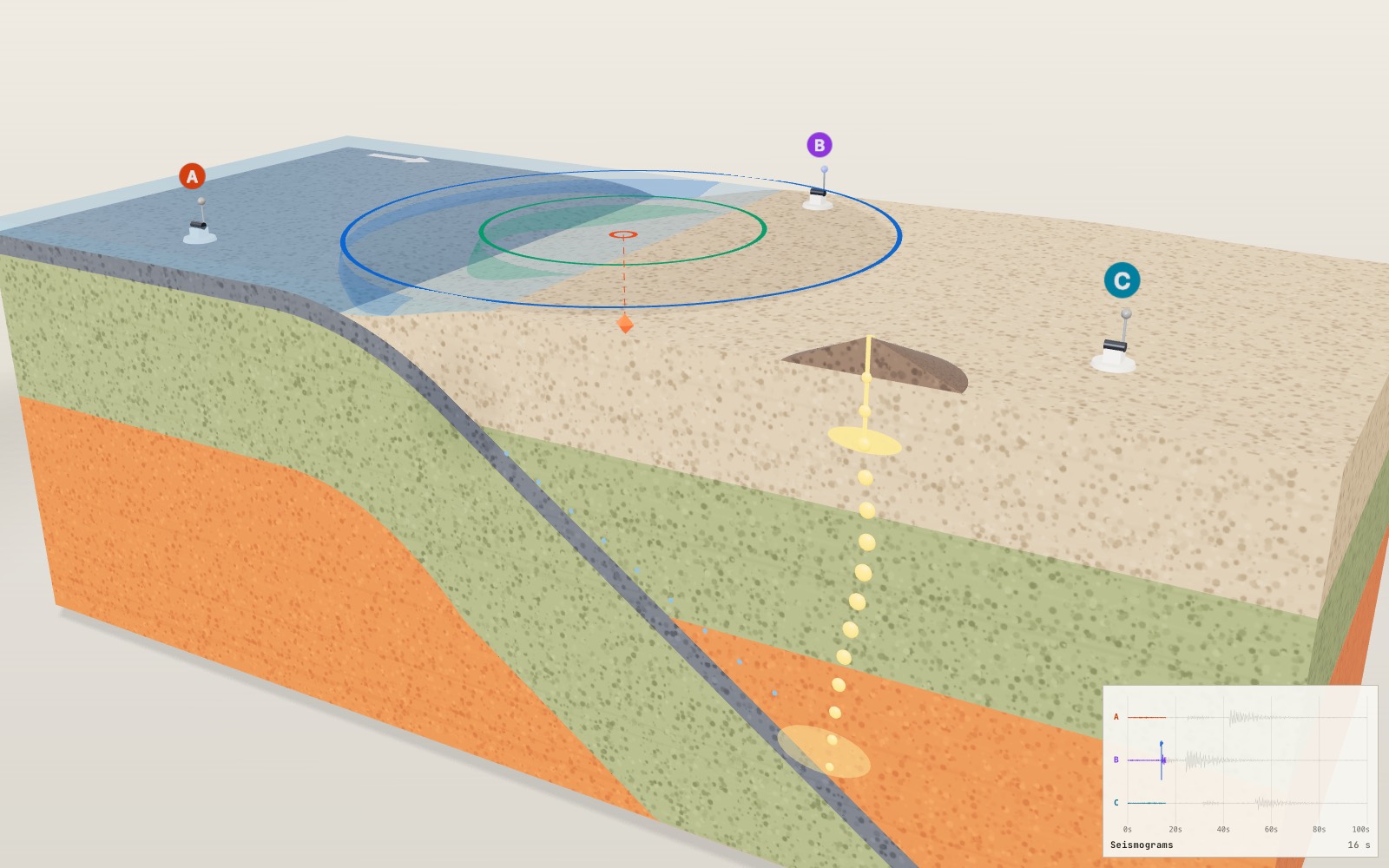
Where volcanoes and earthquakes happen
For each second between the P wave and the S wave, the earthquake is roughly 8 km farther from the seismometer.
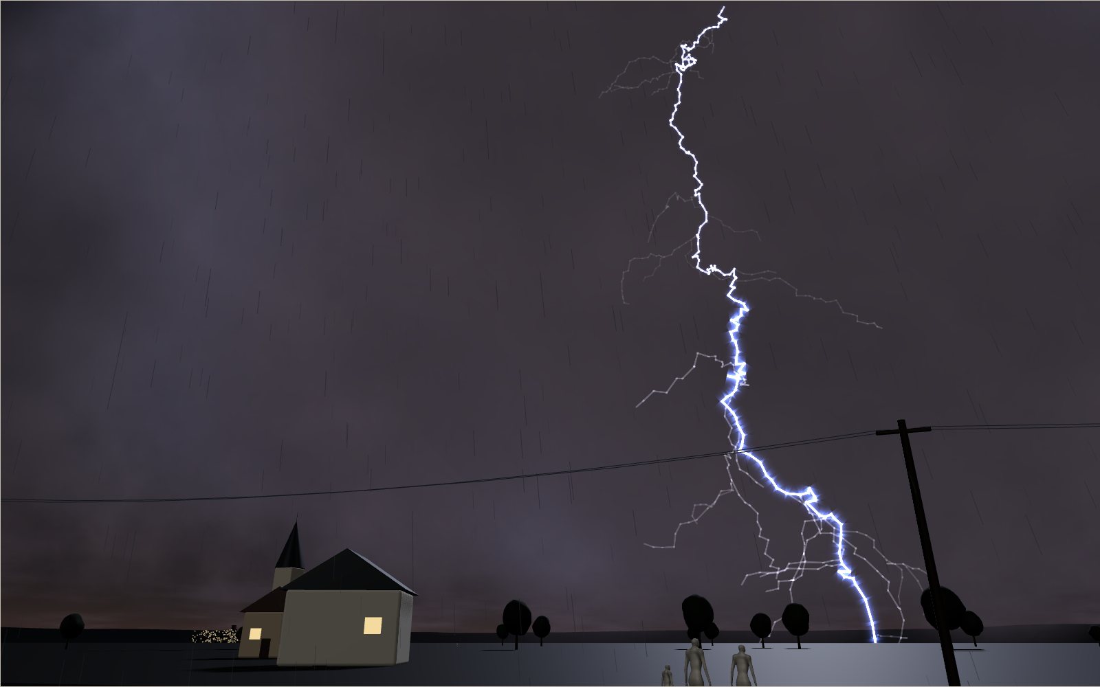
How lightning and thunder work
A lightning channel heats the air to about five times the temperature of the Sun’s surface, and its thunder needs about 3 seconds to cover each kilometre to you.
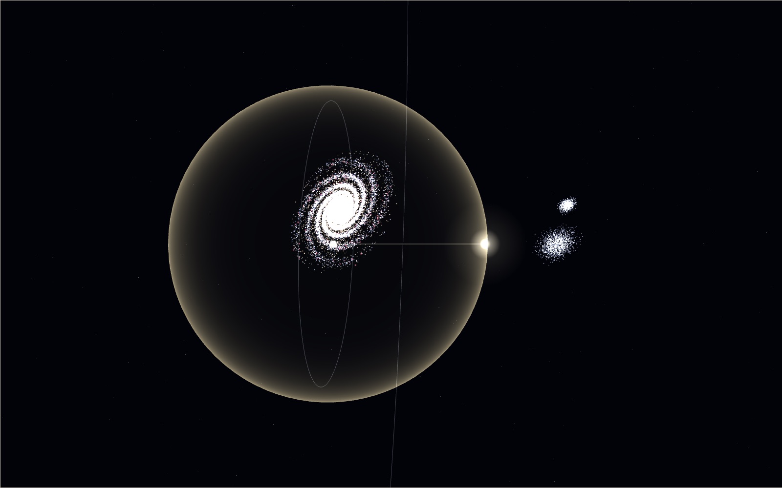
Travelling at the speed of light
Light goes round the equator 7.5 times in a second, yet the same light needs 2.5 million years to reach the Andromeda galaxy.
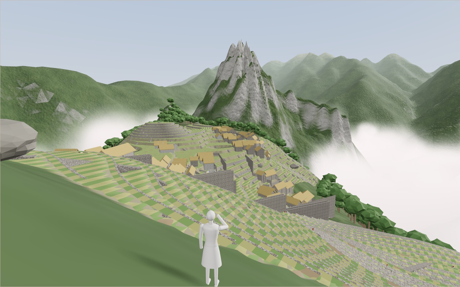
Walk through Machu Picchu
Most of the work that holds Machu Picchu on its ridge, 60 per cent by one estimate, lies under the ground in foundations and drains nobody sees.
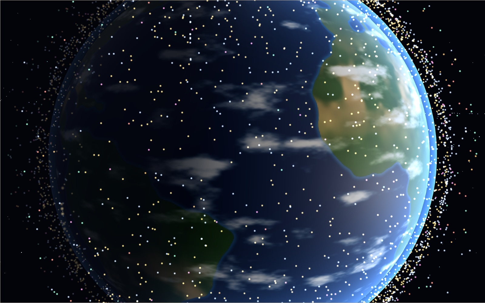
Satellites orbiting Earth right now
On 20 October 2026 the public catalogue listed 16,768 working satellites, and two in every three belonged to one company's internet network.
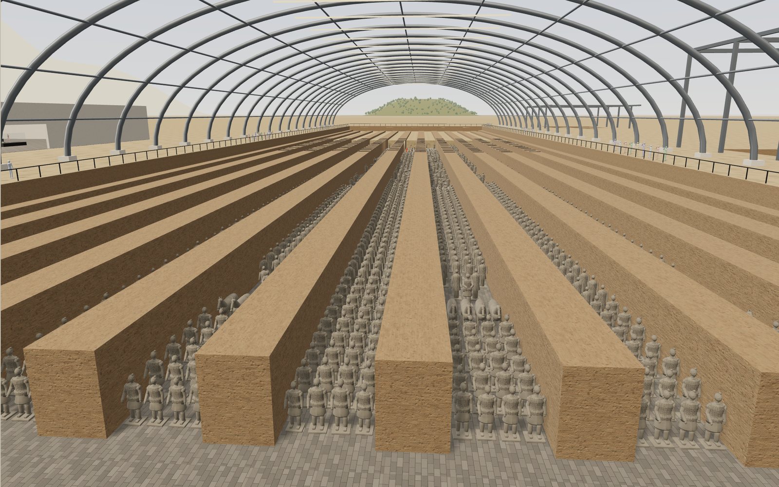
Walk through the Terracotta Army
Pit 1 of the Terracotta Army is 230 m long and holds an estimated 6,000 figures and horses, and every one of them was painted once.
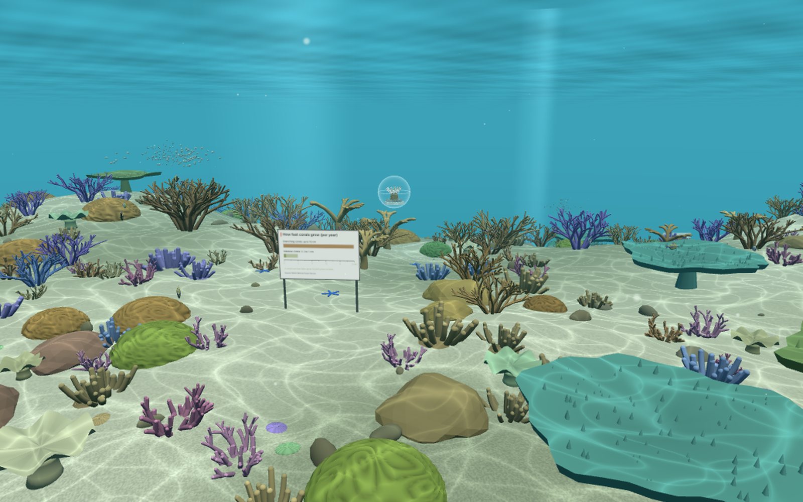
Coral bleaching causes: why warmer water turns reefs white
Corals get most of their food from algae living in their tissue, and when the sea stays more than 1 °C above its usual summer high for weeks, they lose those algae and turn white.
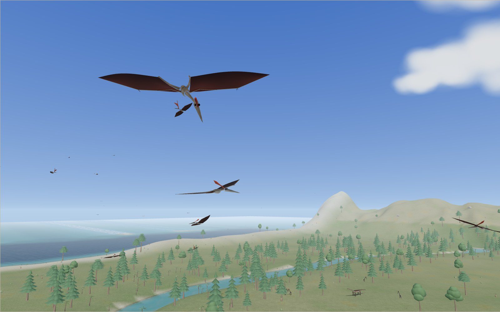
Fly with life-size pterosaurs
Quetzalcoatlus spanned about 10 m, close to the 11 m wing of a Cessna 172, and standing it was as tall as a giraffe. Watch it pass over a Cretaceous cliff, then fly with 35 pterosaurs from three ages.